
Page 63 - 2019_01-Haematologica-web
P. 63
Stromal cells need macrophage iron
A
BC
D
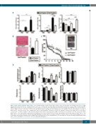
Figure 4. Skin repair is delayed in Fpn1fl/flLysCre+/- mice. (A) Macrophages (CD45+/CD11b+/Ly6C+/F4/80+) were sorted by FACS from wounded skin tissue of eight animals/group from Fpn1fl/flLysCre-/- and Fpn1fl/flLysCre+/- mice at 2, 7 and 12 days post-injury (dpi). Ferroportin (FPN), transferrin receptor 1 (TfR1) and H-ferritin (FtH) mRNA expression was assessed by quantitative real time polymerase chain reaction and normalized to the housekeeping gene 18S RNA. Data are presented as mean ± SEM; *P<0.01, **P<0.001, ***P<0.0001. (B) Representative histology of Perls' Prussian blue iron staining of dorsal skin samples at 12 dpi in Fpn1fl/flLysCre-/- and Fpn1fl/flLysCre+/-mice. Magnification 40X. A semi-quantitative evaluation of Perls' iron staining is shown on the right; n=6 for each group; ***P<0.0001. (C) Kinetic analysis of skin excisional wound areas. Values represent mean ± SEM of 24 values for each group; *P<0.01, **P<0.001 versus Fpn1fl/flLysCre-/-. One representative experiment (6 mice/group) out of four is shown. The inset shows representative macroscopic images of Fpn1fl/flLysCre-/- and Fpn1fl/flLysCre+/- mice skin wounds at 7 dpi. (D) Histological grading of wounds, based on separate evaluation of distinct features of the wound healing process, at 2, 7 and 12 dpi. IGT: immature granulation tissue, MGT: mature granulation tissue. The semiquantitative score was defined as described in Online Supplementary Table S2; n= 12 for each group; ***P<0.0001, **P<0.001 versus Fpn1fl/flLysCre-/-.
haematologica | 2019; 104(1)
53
