
Page 65 - 2019_01-Haematologica-web
P. 65
Stromal cells need macrophage iron
wound lysates, but did not detect significant differences between the two mouse lines at any time-point (Online Supplementary Figure S5). Since iron accumulation in macrophages of Fpn1fl/flLysCre+/- mice could affect the polarization of these cells during the healing process,15 we investigated the distribution of the different polarized macrophages. As expected, an increase in
MHCII+/CD206- M1 macrophages was detected already in the middle-proliferative phase, while a significant increase in MHCII-/CD206+ M2 macrophages was evi- dent only in the late-remodeling phase, but no difference was found between Fpn1fl/flLysCre+/- mice and their con- trol littermates (Figure 5A). Moreover, we evaluated the expression of polarization markers in bone marrow-
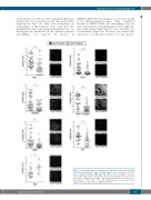
Figure 6. Vessel and stromal cell reduction accompanied by iron deficiency and decreased proliferation in wounds of Fpn1fl/flLysCre+/- mice. Expression of CD31, Lyve-1, collagen-1, PDFGR, αSMA, Ki67 and TfR1 after skin wounding at 7 dpi was assessed by confocal microscopy and the positive area expressed as %. Each circle represents an analysis from a single confocal image (5-9 fields of vision/mouse, 6 mice/group), ***P<0.0001, **P<0.001. Representative confocal microscopy images are shown. Bars: 100 mm. Magnification: 40X.
haematologica | 2019; 104(1)
55
