
Page 62 - 2019_01-Haematologica-web
P. 62
S. Recalcati et al.
Neutrophils (Ly6G+ cells) and eosinophils (CCR3+ cells) were abundant at 2 and 7 dpi and decreased thereafter, whereas an inverse trend was evident for T cells (CD3+ cells) and macrophages (F4/80+ cells), which increased at 12 dpi (Figure 5A). The accumulation kinetics of these cells, which are typical of skin wound healing,22 were not affect-
ed by the presence or absence of FPN in macrophages. Macrophages with different functional orientations have specific roles in the overlapping phases of wound repair.1,9 As iron accumulation in macrophages might favor the expression of inflammatory mediators.17,26,28,29 we evaluated the levels of inflammatory cytokines in
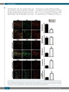
Figure 3. Epithelial iron deficiency and decreased proliferation in cutaneous hair follicles of Fpn1fl/flLysCre+/- mice. Expression and localization of Ki67, F4/80, trans- ferrin receptor (TfR1) and L ferritin subunit (FtL) in cutaneous tissue of Fpn1fl/flLysCre-/- and Fpn1fl/flLysCre+/- mice was assessed by confocal microscopy. Representative confocal microscopy images for merged signals, Ki67, TfR1, F4/80 and FtL are shown. Quantification of confocal images (5-9 fields of vision/mouse, 3 mice/group) is also reported. ***P<0.0001 versus Fpn1fl/flLysCre-/-. Arrowheads indicate hair bulbs. Bars: 100 mm. Magnification: 40X.
52
haematologica | 2019; 104(1)
