
Page 61 - 2019_01-Haematologica-web
P. 61
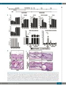
Stromal cells need macrophage iron
A
BC
D
E
Figure 2. Alopecia in Fpn1fl/flLysCre+/- mice is not related to iron deficiency/anemia. (A) Schematic overview of the feeding protocol: pups were fed by dams kept on an iron-deficient diet for 3 weeks until weaning and then maintained on a low iron diet for another 2 weeks followed by a normal diet. (B) Hemoglobin (Hb) levels and hema- tocrit (Hct) in 3- to 8-week old mice (mean ± SEM of 10 mice for each group). The histogram at the bottom shows the degree of alopecia at different time-points. ***P<0.0001 versus Fpn1fl/flLysCre-/-. (C) Top: Hb levels, Hct, serum iron and transferrin saturation (TS) in 3- to 18-week old mice (mean ± SEM of 10 mice for each group; ***P<0.0001). Bottom: hepcidin (HAMP) and erythroferrone (Fam132b) mRNA levels in the liver and spleen, respectively, of 3- to 18-week old mice. Expression in 3- week old mice fed the normal diet is shown in comparison. mRNA levels were measured by quantitative real-time polymerase chain reaction and normalized to the house- keeping gene 18S RNA. Data are presented as mean ± SEM of 10 mice for each group; ***P<0.0001, **P<0.001. (D) Representative histology of the skin (dorsal area) of 3-week old Fpn1fl/flLysCre-/- and Fpn1fl/flLysCre+/- mice maintained on an iron-deficient diet. Magnification 20X. (E) Representative histology of the skin (dorsal region) after 5 weeks of an iron deficient diet plus 2 weeks of a normal diet. Tissue sections were stained with hematoxylin and eosin. Magnification: 10X; 20X in the insets.
haematologica | 2019; 104(1)
51
