
Page 50 - 2019_01-Haematologica-web
P. 50
D. Ingenhag et al.
all decrease in T cells (Figure 5A), valid for both CD4+ and CD8+ T cells (Figure 5B), while the frequency of B cells varied. Addressing the myeloid lineage, an increased fre- quency of monocytes and granulocytes was observed (Figure 5A). Overall blood cellularity was decreased in B6[HB9] regardless of lineage contribution (Figure 5C). Serial blood draws confirmed these observations during the entire monitoring period (6.5 months). To investigate whether a differentiation blockage is causative for reduced blood cellularity as well as absence of HB9-GFP+ mature blood cells, we performed comprehensive FACS analysis from HSCs to mature B cells, T cells, and myeloid cells.
In contrast to the mature cell pool, HB9-GFP-expressing cells were detected within the Lin–Sca-1+c-Kit+ (LSK)/HSC compartment (Figure 6A). To evaluate the differentiation potential of HB9-expressing HSCs, Lin– cells were further analyzed regarding IL7Rα, which is expressed by lym- phoid but not myeloid committed progenitors. HB9-GFP expression was decreased in lymphoid committed progen- itors (Lin–IL7Rα+) compared to that in myeloid committed progenitors (Lin–IL7Rα–), while B6[GFP] showed compara- ble amounts of GFP+ cells in both progenitor populations (Figure 6B). Due to the enrichment of HB9-expressing cells in the myeloid progenitor subset, this compartment was further analyzed. Sub-gating of the myeloid progenitor population into common myeloid progenitors (CMP), megakaryocyte/erythrocyte progenitors (MEP) and granu- locyte/macrophage progenitors (GMP) revealed that HB9- GFP-expressing cells contributed almost exclusively to the MEP subset as the amount of HB9-GFP+ MEP was signifi-
cantly enriched compared to CMP and GMP (Figure 6C). In line with peripheral blood, total white blood cell number of bone marrow and thus CMP, GMP and MEP was decreased in B6[HB9] compared to B6[GFP], but with- out affecting cell frequencies (Online Supplementary Figure
S6).
Analysis of terminal MEP maturation was not applica-
ble, as erythrocytes as well as thrombocytes are anuclear cell types and therefore do not carry the transgene. We further assessed the differentiation potential of HB9-GFP+ GMP cells, together with the frequency of the myeloid cell types originating from this. According to peripheral blood analysis, no distinct HB9-GFP+ cell population was detectable within the granulocyte and the monocyte/macrophage population in the bone marrow or spleen of B6[HB9] compared to B6[GFP] (Online Supplementary Figures S7-S9 and S11A-C). The frequency of macrophages/monocytes was slightly increased in B6[HB9] compared to B6[GFP], which is congruent to peripheral blood analysis, while, in contrast, the frequen- cy of granulocytes was comparable (Online Supplementary Figure S12A and B).
With respect to lymphoid differentiation we assessed HB9-GFP expression as well as frequency of immature and mature B cells and T cells. While at the immature B220+CD19+CD93+ B-cell stage a low frequency of HB9- GFP+ cells was still detectable, no distinct HB9-GFP+ cell population was identified within the more mature B220+CD19+CD93– B-cell population in bone marrow, or within mature B cells of the spleen and peripheral blood
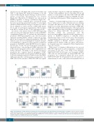
AB
C
Figure 4. Transplantation of HB9-transduced hematopoietic stem and progenitor cells and monitoring of hematopoietic reconstitution. (A) Flow cytometric analysis of GFP expression in Lineage– cells, non-transduced (Lin[nt]), or transduced with HB9 (Lin[HB9]) or control vector (Lin[GFP]). Shown is one representative experiment. (B) Detection of HB9 expression by qRT-PCR in Lin[GFP] and Lin[HB9] (n=3). (C) Flow cytometric analysis of CD45.2 and GFP expression in peripheral blood cells of Lin[GFP]- (B6[GFP]) or Lin[HB9]-transplanted mice (B6[HB9]).
40
haematologica | 2019; 104(1)
