
Page 52 - 2019_01-Haematologica-web
P. 52
D. Ingenhag et al.
CD34[HB9] cells displayed transgene expression levels (Figure 7A) comparable to those in translocation t(7;12) AML blast cells,22 and no endogenous HB9 expression was detectable (Figure 7B).
Genome-wide expression profiling revealed 117 genes, which were significantly differentially expressed (P≤0.05, FC≥1.8) in CD34[HB9] compared to CD34[GFP] (Online
Supplementary Table S1 and Online Supplementary Figure S13). The gene with the highest fold change was HB9/MNX1, confirming transgene expression. Six genes were de novo expressed in CD34[HB9] compared to CD34[GFP] (Figure 7C), of which hemoglobin subunit zeta (HBZ), as well as solute carrier family 4 member 1 (SLC4A1), are restricted to the erythroid lineage, thereby
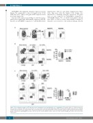
A
B
C
Figure 6. Flow cytometric analysis of GFP expression in the hematopoietic stem and progenitor cell compartment. (A) Lineage–c-Kit+Sca-1+ (LSK) hematopoietic stem cells were analyzed for GFP expression. Shown is one representative experiment. (B) Lymphoid (Lin–IL7Rα+) and myeloid (Lin–IL7Rα–) progenitors were analyzed for GFP expression and the percentage of GFP+ cells was determined for each compartment (n=5). (C) Flow cytometric analysis of GFP expression in the myeloid pro- genitor compartment, regarding MEP (Lin- IL7Rα– c-Kit+ Sca-1- FcγRlow CD34-), CMP (Lin– IL7Rα- c-Kit+ Sca-1- FcγRlow CD34+) and GMP (Lin– IL7Rα– c-Kit+ Sca-1– FcγRhigh CD34+) (n=5).
42
haematologica | 2019; 104(1)
