
Page 51 - 2019_01-Haematologica-web
P. 51
HB9 induces senescence
(Online Supplementary Figure S7-S9 and S11D and E). Furthermore, B6[HB9] mice showed an overall increased frequency of immature CD93+ and a decreased frequency of more mature CD93– B cells in bone marrow compared to B6[GFP], but without affecting B-cell frequency in the periphery (Online Supplementary Figure S12C).
In contrast to immature B cells, no HB9-GFP+ cells were detected within immature CD4+CD8+ thymocytes (Online Supplementary Figures S10 and S11F). Congruent to the other lineages, no distinct HB9-GFP+ cell population was found within the mature T-cell compartment of bone mar- row, spleen or peripheral blood (Online Supplementary Figure S7-S9 and S11G). In line with our results obtained from peripheral blood, the frequency of CD8+ T cells was slightly decreased in bone marrow and spleen of B6[HB9] compared to B6[GFP], while the frequency of CD4+ T cells was comparable (Online Supplementary Figure S12D). In
summary, HB9+ HSCs showed an impaired differentiation capacity, resulting in an overall differentiation blockage, leading to reduced bone marrow as well as peripheral blood cellularity and accumulation of HB9+ cells at the MEP stage.
HB9 triggers expression of erythropoiesis-related genes and leads to decreased clonogenic potential in HSPCs
To further analyze the influence of HB9 on hematopoi- etic cells, regarding gene expression as well as clonogenic- ity, a highly purified population of transduced cells is mandatory. Thus, these experiments were performed with primary human CD34+ HSPCs, as these cells display a better GFP signal-to-noise ratio compared to their murine counterpart, allowing GFP+ cell FACS-sort upon transduction.
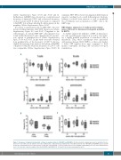
A
B
C
Figure 5. Frequency of lymphoid and myeloid cell types in peripheral blood of B6[GFP] and B6[HB9]. (A) Flow cytometric analysis was used to determine the fre- quency of T cells (CD3+), B cells (CD19+), monocytes (CD11b+Gr-1—) and granulocytes (CD11b+Gr-1+) in peripheral blood cells of B6[GFP] and B6[HB9] 12, 19 and 26 weeks after transplantation (n=6). (B) Frequency of CD3+CD4+ and CD3+CD8+ T cells in peripheral blood cells of B6[GFP] and B6[HB9] 26 weeks after transplantation (n=6). (C) Leukocyte count/mL peripheral blood of B6[GFP] and B6[HB9] 12, 19 and 26 weeks after transplantation (n=6).
haematologica | 2019; 104(1)
41
