
Page 69 - 2018_11-Haematologica-web
P. 69
Obtaining blood stem cells for gene transfer in FA
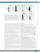
Figure 1. Diminished CD34Hi hematopoietic cells from Fanconi Anemia A genetic defect (FA-A) patients. CD34 expression in baseline bone marrow (BM) (Patients 1, 2, 3, and healthy donor 1) or mobilized leukapheresis (mAPH) (Patient 3 and healthy donor 2) products was determined by fluores- cence staining and flow cytometry analysis. Positive cell fractions are gated based on unstained and isotype stained control samples into two levels of CD34 expression: low expression, CD34Lo, or high expression, CD34Hi. The average mean fluorescence intensity (MFI) of CD34Lo population = 3453; standard error of the mean (SEM) = 516 and CD34Hi population = 19731; SEM = 4103.
reverse, 5'-CCGTGCGCGCTTCAG; probe, 5'-AGCTCTCTC- GACGCAGGACTCGGC (Integrated DNA Technologies; IDT, Coralville, IA, USA)] and in a separate reaction with a β-globin- specific primer/probe combination [forward, 5’-CCTATCA- GAAAGTGGTGGCTGG; reverse, 5'-TTGGACAGCAA- GAAAGTGAGCTT; probe, 5'-TGGCTAATGCCCTGGCCCA- CAAGTA (IDT)]. Two standard curves were established by seri- al dilution of gDNA isolated from a human cell line (HT1080) confirmed to contain a single integrant of the same LV backbone and from peripheral leukocytes collected from a healthy donor using both primer-probe sets independently.
Individual colony gDNA samples were subjected to multiplex real-time TaqMan qPCR to amplify the LV-specific product and an endogenous control (TaqMan Copy Number Reference assay RNaseP, Thermo Fisher Scientific, Pittsburgh, PA, USA). Samples with an average VCN ≥0.5 were considered transduced.
Flow cytometry analysis of hematopoietic subsets
Stained cells were acquired on a FACSCantoTM II, FACSAriaTM II or FACS LSR II (all from BD Bioscience) and analyzed using FlowJo software v.10.0.8 (Tree Star Inc., Ashland, OR, USA). Analysis was performed on up to 20,000 cells. Gates were estab- lished using Full Minus One stained controls.
Antibodies included anti-human CD34 (clone 563), CD16 (clone 3G8), CD3 (clone UCHT1), CD4 (clone L200), CD8 (clone RPA-T8), all from BD Biosciences; CD14 (clone 61D3, Thermo Fisher Scientific, Pittsburgh, PA, USA); CD19 (clone 4G7, BD Pharmingen, San Diego, CA, USA); CD90 (clone 5E10), CD20 (clone 2H7), CD15 (clone W6D3), all from Biolegend (San Diego, CA, USA); CD133 (clone 293C3, Miltenyi Biotec, GmbH); CD45 (clone D058-1283) and CD45RA (clone 5H9), both from BD Horizon (San Jose, CA, USA).
For mouse samples, antibodies were anti-mouse CD45-V500 (561487, clone 30-F11), anti-human CD45-PerCP (347464, clone 2D1), CD3-FITC (555332, clone UCHT1), CD4-V450 (560345, clone RPA-T4), CD8-APCCy7 (557834, clone SK1), CD20-PE (555623, clone 2H7), and CD14-APC (555824, clone 581), all from BD Biosciences.
Results
Diminished CD34Hi expressing cells in FA-A BM and mAPH
Two enrolled patients underwent BM harvest to collect available CD34+ HSPCs (Patients 1 and 2). The third patient underwent mobilization with filgrastim and pler- ixafor followed by peripheral blood leukapheresis (Patient 3). All 3 patients demonstrated reduced CD34 expression and estimated numbers of CD34+ cells in screening BM aspirate samples prior to collection and treatment, relative to healthy donor BM products, as well as in cell products collected for CD34+ cell isolation and gene transfer (Figure 1). Two levels of CD34 expression were observed, CD34Lo [mean fluorescence intensity (MFI)=3453±516], and CD34Hi (MFI=19731±4103). Notably, the proportion of CD34Hi cells were markedly reduced in FA-A patients relative to those observed in healthy donors (Figure 1).
FA-A CD34Hi cells, but not CD34Lo cells, demonstrate in vitro repopulating capacity
To determine which CD34+ cells demonstrated repopu- lation potential, we used colony-forming cell (CFC) poten- tial as a surrogate. This required sufficient blood product to flow-sort CD34Lo and CD34Hi cells for in vitro assays. Only the mAPH product collected from Patient 3 was suf- ficient for this study. For direct comparison, we sort-puri- fied CD34Lo and CD34Hi cells from a healthy donor mAPH product. Only CD34Hi cells from the FA-A patient demon- strated colony-forming potential (Figure 2A). In the healthy donor, CD34Hi cells also demonstrated the major- ity of CFC capacity in comparison with CD34Lo cells, and at much higher levels as compared to the FA-A patient (Figure 2B). These data suggest repopulating capacity is restricted to CD34Hi cell fractions, underscoring the need to preserve as many of these cells as possible for gene transfer processes.
haematologica | 2018; 103(11)
1809
