
Page 68 - 2018_11-Haematologica-web
P. 68
J.E. Adair et al.
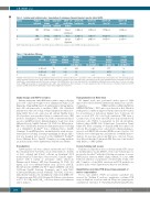
Table 2. Isolation and lentiviral vector transduction of autologous Fanconi Anemia A genetic defect HSPC.
Patient
1
2 3
Product Product collected volume
processed
BM 1278 mL
BM 534 mL mAPH 513 mL
Total CD34+ cells collected
1.69E+08
1.59E+07 9.5E+07
CD34+ cell purification method
Direct
enrichment
None
Lineage
depletion
CD34+ cells transduced
5.34E+06
8.15E+06 5.28E+07
CD34+ cell dose/kg
3.57E+04
3.57E+05 2.44E+06
CD34Hi cell dose/kg
1.77E+04
4.62E+04 3.69E+05
% Gene transfer in CFCs,
CD34Lo cell dose/kg
1.65E+04
3.15E+05 2.06E+06
% Gene transfer in CFCs,
CD34Hi: high CD34 expression; CD34Lo: low CD34 expression; BM: bone marrow product; mAPH: mobilized apheresis product.
Table 3. Transduction efficiency.
Patient
1
2
MOI (IU/cell)
10 IU/ cell
10 IU/ cell
Viability of infusion product (%)
79
99.5
VCN of infusion product
0.33
1.83
Plating efficiency in CFCs,
Plating efficiency in CFCs,
0nMMMC(%) 10nMMMC(%) 0nMMMC(%) 10nMMMC(%
4.14 0.22 17.7 80
3.8 0.03 42.73 100
3 5IU/cell 99.3 0.67 2.75 0.04 26.23 100
Viability of the infusion product was determined by trypan blue dye exclusion.The vector copy number (VCN) in the bulk transduced population was determined by quanti- tative polymerase chain reaction (qPCR) method against a reference standard curve.Plating efficiency of the infusion product was determined as the percentage of CD34+ cells plated with colony-forming capacity. Functional correction of the FANCA gene defect was determined by calculating the plating efficiency under stress of various concentra- tions of MMC. Gene transfer in colony-forming cells (CFCs) was determined as the percentage of colonies analyzed positive for the presence of lentivirus backbone by PCR analysis on DNA extracted from individual colonies. MOI: multiplicity of infection.
Study design and HSPC isolation
Patients underwent either BM harvest with a target collection goal of 15 cc/kg body weight or were administered daily G-CSF (filgrastim; 16 mg/kg BID; days 1-6) and plerixafor (240 mg/kg/day; days 4-6) subcutaneously to mobilize CD34+ cells. Mobilized patients were subjected to large volume leukapheresis when circu- lating CD34+ blood cell counts were ≥5 cells/mL. Healthy donor blood products were purchased from a commercial source (BM products; StemExpress, Folsom, CA, USA) or institutional shared resources (mAPH products). Immunomagnetic beads were from Miltenyi Biotech, GmbH (Auburn, CA, USA). For BM products, RBC were debulked by hetastarch sedimentation prior to labeling on a CliniMACS ProdigyTM device (Miltenyi Biotec GmbH, Germany). For mAPH products, an initial platelet wash was per- formed prior to labeling. Custom programming for lineage deple- tion was designed and executed on the CliniMACS ProdigyTM device (Miltenyi Biotec, GmbH). Complete processing methods are included in the Online Supplementary Materials and Methods.
Transduction
Transplantation in NSG mice
All animal work was performed under protocol 1864 approved by the Fred Hutch Institutional Animal Care and Use Committee. NOD.Cg-PrkdcscidIL2rγtmlWj/Szj (NOD/SCID/IL2rγnull, NSG) mice were housed at Fred Hutch in pathogen-free conditions approved by the American Association for Accreditation of Laboratory Animal Care. 8-12-week old mice received 275 cGy total body irradiation (TBI) from a Cesium source. Four hours after TBI, 1x10⁶ gene-modified total nucleated cells (TNCs) re-suspended in 200 mL phosphate buffered saline (D-PBS, Life Technologies Corporation, Grand Island, NY, USA) containing 1% heparin (APP) were infused via tail vein. Blood samples were collected into ethylenediaminete- traacetic acid (EDTA) Microtainers (BD Bioscience, San Jose, CA, USA) by retro-orbital puncture and diluted 1:1 with PBS prior to analysis. At necropsy, spleen and BM were collected. Tissues were filtered through 70 mm mesh (BD Bioscience) and washed with Dulbecco’s PBS (D-PBS).
Colony-forming cell assays
Transduced cell products were seeded in standard CFC assays in methylcellulose media (H4230, Stem Cell Technologies) as previously described16 with the following exceptions: to assess FANCA gene function, MMC (Sigma Aldrich, St. Louis, MO, USA) was added at concentrations of 0 nM, 5 nM, 10 nM, or 20 nM. Complete colony DNA extraction and PCR methods are included in the Online Supplementary Materials and Methods.
Quantitative real-time PCR-based measurement of vector copy number
Vector copy number (VCN) per genome equivalent was assessed by TaqMan 5' nuclease quantitative real-time PCR assay in duplicate reactions with an LV-specific primer/probe combination [forward, 5'-TGAAAGCGAAAGGGAAACCA;
CD34-enriched cells were cultured on RetroNectin (Takara Bio, Mountain View, CA, USA)-coated culture flasks at a density of 1x10⁶ cells/mL and 2.9x10⁵ cells/cm2 in StemSpanTM ACF media (StemCell Technologies, Vancouver, BC, Canada), supple- mented with 4 mg/mL of protamine sulfate (American Pharmaceutical Partners; APP, East Shaumburg, IL, USA), 100 ng/mL each of recombinant human stem cell factor (rhSCF), thrombopoietin (rhTPO) and Flt-3 ligand (rhFLT3L) (all from CellGenix GmbH, Freiburg, Germany), and 1 mM NAC (Cumberland Pharmaceuticals, Nashville, TN, USA). Cells were immediately transduced at a multiplicity of infection (MOI) of 5- 10 infectious units (IU)/cell. Following 12-24 hours of incubation at 37°C, 5% CO2 and 5% O2 , cells were harvested for infusion and/or analyses.
1808
haematologica | 2018; 103(11)
