
Page 70 - 2018_11-Haematologica-web
P. 70
J.E. Adair et al.
Extensive loss of FA-A CD34Hi cells with direct clinical purification protocols
The current clinical standard for CD34+ cell enrichment is optimized for collection of CD34Hi cells. However, in Patient 1, direct enrichment of CD34+ cells using this pro- tocol was inefficient, resulting in an approximately 3% yield and only 5.34x10⁶ total CD34+ cells available for gene transfer (Table 2). Moreover, the purity of the enriched cell product was only 58.9%, and approximately 47% loss in viable cells was observed during culture and gene transfer. Resulting gene-modified cells retained colony-forming capacity and demonstrated acquired resistance to the potent DNA crosslinking agent MMC following LV-medi- ated FANCA gene transfer (Table 3).
In Patient 2, estimated losses during direct CD34 enrich- ment and gene transfer were expected to reduce the cell product available for transduction to a level lower than observed for Patient 1. Thus, an urgent amendment was filed with the FDA to permit elimination of the direct CD34 enrichment steps and allow transduction of the entire red blood cell (RBC)-depleted BM product. This processing change preserved more CD34+ cells (Table 2), with improved transduction and viability (Table 3). Together, these data suggested that minimal manipulation of target CD34+ cells from FA-A patients could improve yield, gene transfer efficiency, and function in vivo.
Development of a novel strategy to deplete lineage+ cells
We hypothesized that depleting non-target mature B cells, T cells, monocytes, and granulocytes would retain precious CD34+ cells with minimal manipulation, since CD34-expressing cells would not be directly labeled, selected, or washed (Figure 3). Building on our previous work automating cell selection and gene transfer using the CliniMACS ProdigyTM device,17 we designed a cus- tomized, automated RBC debulking and immunomagnet- ic bead-based lineage specific depletion strategy (Online Supplementary Materials and Methods). Four different bead- conjugated antibody reagents were used in this approach: anti-CD3 (T-cell removal), anti-CD14 (monocyte removal), anti-CD16 (granulocyte and NK-cell removal), and anti-CD19 (B-cell removal). This protocol was designed for both BM and mAPH products.
Lineage depletion preserves available CD34+ cells for gene transfer
A total of nine BM and ten mAPH products were processed to establish process validity. An average 60% of BM CD45+ cells and 50% of mAPH CD45+ cells expressed one of the four target markers (CD3, CD14, CD16, or CD19) (Online Supplementary Figure S1A and B, respective- ly). CD34+ cell content in these products ranged from 0.35- 1.4% in BM and 0.06-0.9% in mAPH products. The aver- age process run time for BM products was ten hours, whereas mAPH products were processed over 13 hours. Observed total nucleated cell (TNC) reduction was approximately 1 log for both BM and mAPH products fol- lowing lineage depletion (Figure 4A). All target lineage+ cells were depleted to less than 10% of initial numbers, and CD34+ cells were retained at 94.62±4.61% for BM products and 70.69±11.4% for mAPH products (Figure 4B). Retention of available CD34Hi and CD34Lo cells was observed and comparable or superior to that observed for the same products by direct CD34-enrichment (Online
Supplementary Figure S2). Approximately 24% of BM CD34+ cells were colony-forming in a standard methylcel- lulose assay, while 51% of mAPH CD34+ cells formed colonies (Figure 4C and Online Supplementary Figure S3). However, following LV transduction of these cells using the same protocol proposed for FA-A patient cells, we observed consistent 50% rates of gene transfer into CFCs from both cell product types (Figure 4D). Analysis of sin- gle colonies demonstrated an average VCN per CFC of 0.7 for BM CD34+ cells and 1.6 for mAPH CD34+ cells. VCN was also assessed in bulk transduced cells cultured for ten days in vitro, demonstrating an average value of 5 for both BM and mAPH products (Figure 4E). Final cell products tested for mycoplasma and sterility were negative, and endotoxin testing demonstrated values within criteria for patient infusion. Lineage-depleted and transduced cells from six mAPH and BM products each were infused into immunodeficient (NSG) mice at a target cell dose of 1x10⁶ TNC per mouse. On average, the CD34+ cell dose per mouse for BM products was 2.86x10⁴ CD34+ cells [stan- dard error of the mean (SEM)=6.67x10³] and for mAPH products was 1.08x10⁵ CD34+ cells (SEM=1.45×10⁴). Flow cytometry analysis on peripheral blood was used to eval- uate engraftment (human CD45+) and lineage develop- ment into T cells (human CD3+), B cells (human CD20+),
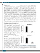
Figure 2. In vitro repopulation potential restricted to CD34Hi hematopoietic cells. Mobilized leukapheresis from FA-A Patient 3 (Panel A) and a healthy donor (Panel B) were in parallel fluorescence stained with anti-CD34 antibody and sort-purified for CD34Hi and CD34Lo cells. Total nucleated cells (TNC) equivalent to 1500 CD34-expressing cells were seeded in CFC assays. Percentage of CD34+ cells seeded in the assay that gave rise to colonies is represented as the % of colony-forming cells.
A
B
1810
haematologica | 2018; 103(11)
