
Page 71 - 2018_11-Haematologica-web
P. 71
Obtaining blood stem cells for gene transfer in FA
and monocytes (human CD14+) over time (Figure 4F). Both mAPH and BM products demonstrated long-term engraftment over 20 weeks of monitoring. Engraftment levels were comparable to results reported by Wiekmeijer et al. with CD34+ cells purified from BM and infused at similar cell doses.18
Lineage-depleted cell products xenoengraft equivalently to CD34-enriched products
In this experiment, healthy donor BM products were divided into two aliquots. One was lineage-depleted and the other CD34-enriched. Resulting cell populations were transduced with the same LV vector under identical condi- tions and infused into NSG mice at matched CD34+ cell doses. We observed higher CD34+ cell retention with lin- eage depletion compared to CD34 selection, with no dif- ferences in transduction efficiency or colony-forming potential (Figure 5A and B). We observed slightly higher, but not significantly different, levels of human CD45+ blood cell engraftment in mice receiving transduced, line- age-depleted cells relative to mice receiving CD34-select- ed cells. We also observed more stability of T- and B-cell engraftment in mice receiving lineage-depleted cell prod- ucts relative to mice receiving CD34-selected cell products (Figure 5C).
Lineage depletion protocol preserves limited FA CD34Hi cells
These data collectively suggest that lineage-specific depletion preserved available CD34+ cells without com- promising transduction efficiency or cell fitness. Under
FDA approval, the clinical protocol was modified to include both BM and/or mAPH products, with lineage depletion as the method of CD34+ cell enrichment. Patient 3 (the first treated under the modified protocol) was a 5- year old male with FA-A confirmed by complementation studies. Baseline neutrophils averaged 1.7x10⁹/L and base- line platelets averaged 32x109/L in the six months prior to treatment, with declining neutrophils and platelets over the prior 2-year interval (Online Supplementary Figure S4). Mobilization of ≥10 CD34+ cells/mL peripheral blood was achieved (Online Supplementary Figure S5A), and two suc- cessive apheresis collections resulted in 8.5x10¹⁰ TNC con- taining a total 1.6x10⁸ CD34+ cells (Table 2). The patient required a total of two platelet transfusions and two packed red blood cell transfusions during mobilization and leukapheresis (Online Supplementary Figure S5B). Due to column limitations, 5x10¹⁰ TNC (equivalent to 9.5x10⁷ total CD34+ cells) were subjected to lineage depletion, and the remainder were cryopreserved. Lineage depletion resulted in a 94% reduction in TNC and a 56% retention of available CD34+ cells. CD34 purity was 1.6%, repre- senting a 1-2 log-fold increase in the total number of CD34+ cells per kg available for transduction and infusion relative to Patients 1 and 2 (Table 2). A total of 52.8x10⁶ CD34+ cells were transduced at an MOI of 5 IU/cell, resulting in a final cell dose of 2.4x10⁶ total CD34+ cells per kg with 99.3% viability based on trypan blue dye exclu- sion. Approximately 26% of CFCs in this cell product were transduced, displaying a mean VCN of approximate- ly 1 (0.9) (Table 3). Thus, limited numbers of available CD34+ cells were indirectly enriched using lineage deple-
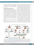
Figure 3. Direct CD34 enrichment versus depletion of lineage positive (+) cells. Products can include bone marrow (BM) or mobilized apheresis product (mAPH) (1). BM products were first processed through hetastarch sedimentation to deplete red blood cells (RBCs). Leukapheresis products were first subjected to several washes to deplete platelets. For direct CD34+ cell selection, anti-CD34 antibody-bound immunomagnetic beads (microbeads) are used, whereas for lineage depletion anti- CD3+, CD14+, CD16+, and CD19+, microbeads are used (2). In both cases, microbead-bound cells are retained on the column and subjected to wash steps. When lin- eage depletion is used, CD34-expressing cells undergo minimal manipulation during purification. Following purification, cells are cultured and transduced with a VSV-G pseudotyped lentiviral vector at a multiplicity of infection (MOI) of 5–10 IU/ cell (3). Following ~16 hours of incubation cells are harvested (4). *These process- es were performed on the CliniMACS ProdigyTM device from Miltenyi Biotec GmbH.
haematologica | 2018; 103(11)
1811
