
Page 67 - 2018_11-Haematologica-web
P. 67
Obtaining blood stem cells for gene transfer in FA
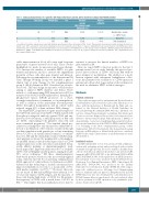
Table 1. Clinical characteristics of 3 patients with Fanconi Anemia A genetic defect enrolled in clinical trial NCT01331018.
Patient
1
2
3
Age (years)
22
10
5
Weight (kg) Sex
70.7 Male
19.6 Male
14.7 Male
Baseline average ANC/platelet count (thousand/mL)
1.3/65
1.0/62
1.7/32
Marrow cellularity
10-30%
~20%
~30%
FANCA defect
Exon22 splice variant
(c. 1827-1 G>A)
Exons6-31
Not determined
One adult and 2 pediatric patients were treated with lentivirus gene therapy for Fanconi Anemia-A (FA-A) defect. Three patients demonstrated steadily declining: absolute neu- trophil count (ANC) and platelet counts in the peripheral blood prior to treatment and less than 30% marrow cellularity. Molecular characterization of the FANCA gene defect performed by gene sequencing demonstrated that Patient 1 was homozygous in the FANCA gene for the splicing variant. For Patient 2, Multiplex Ligation-dependent Probe Amplification (MLBA) on the FANCA gene identified a homozygous gross deletion of exons 6-31.No sequence analysis was performed for Patient 3, but complementation test- ing confirmed FANCA defect.
stable improvements in blood cell counts with long-term persistence of gene-corrected blood cells. These studies highlighted two needs for innovation in FA gene therapy: 1) to increase the number of available HSPCs for gene transfer and infusion; and 2) to increase the engraftment potential of these cells after gene transfer and infusion. Following the recommendations of the International FA Gene Therapy Working Group,8 we launched a phase I clinical trial of gene therapy for FA complementation group A (FA-A) patients in 2011 (clinicaltrials.gov identifier: 01331018). This trial design incorporates several features aiming to improve HSPC numbers and fitness. These include: i) a self-inactivating (SIN) lentiviral vector (LV) for transfer of the FANCA cDNA regulated by a human phos- phoglycerate kinase (hPGK) promoter; ii) a short, overnight transduction to minimize ex vivo manipulation, as well as addition of the antioxidant N-acetylcysteine (NAC) throughout manipulation; and iii) culture under reduced oxygen (5%) to limit oxidative DNA damage.9
The target HSPC population for gene transfer expresses the CD34 cell surface protein (CD34+). When stained with fluorophore-conjugated antibody against CD34 and ana- lyzed by flow cytometry, a small proportion of BM cells are CD34+, representing both primitive stem cells and more committed progenitors.10 The standard clinical pro- cedure for isolating these cells first involves either BM col- lection or mobilization of the cells into circulation through cytokine stimulation with granulocyte colony stimulating factor (G-CSF) or, in certain clinical scenarios, a combina- tion of G-CSF and the chemokine receptor CXCR4 antag- onist plerixafor, followed by peripheral blood leukaphere- sis (mAPH). Initial isolation technologies relied on CD34 antigen expression on the cell surface and utilized biotin- avidin affinity, panning, or immununomagnetic bead- based approaches. Expected yields were 50% of available CD34+ cells with highly variable purities, ranging from 20- 90% across techniques.11 Of these, immunomagnetic bead-based positive selection is the most widely-applied today, with the first US Food and Drug Administration (FDA) approval of a clinical device for human use in 2014. Advances in this technology to include automation have improved reliability in recovery to a mean yield of 70% with purities regularly over 90%.12,13 However, these val- ues are based on BM and mAPH products wherein 1-3% of total cells express CD34 antigen, and the majority of these cells display high levels of CD34. For FA patients, the frequency of CD34+ cells is much lower: 0.1-1.5% in BM.14,15 This implies that non-standard processes may be
required to preserve the limited numbers of HSPCs for gene transfer in FA.
Here we report HSPC collection results for the first 3 patients treated on our study. Initially, this protocol pro- posed direct isolation of CD34+ cells from BM without prior attempts at mobilization. The addition of a mobi- lization regimen with subsequent leukapheresis collec- tions has permitted the evaluation of CD34 expression patterns in both product types and provided evidence for the need for alternative HSPC isolation strategies.
Methods
Patientselection
This study was approved by an Institutional Review Board at Fred Hutchinson Cancer Research Center (Fred Hutch) in accor- dance with the Declaration of Helsinki and the FDA, and con- formed to the National Institutes of Health Guidelines for Research Involving Recombinant DNA Molecules. Informed con- sent was obtained from all patients or guardians. FA patients aged 4 years or over were diagnosed by a positive test for increased sen- sitivity to chromosomal breakage with mitomycin C (MMC) or diepoxybutane. Correction of melphalan hypersensitivity follow- ing retroviral transduction of the FANCA cDNA identified Patient 3 as belonging to the FA-A complementation group. (Online Supplementary Table S1). FA-A patients who demonstrated normal karyotype in BM analyses as defined in the trial were considered eligible for the study. Characteristics of enrolled patients are avail- able in Table 1.
Lentiviral vectors
All SIN lentiviral (LV) vectors were produced with a third-gen- eration split packaging system and pseudotyped with vesicular stomatitis virus glycoprotein. LV used to transduce healthy donor cells encoded either an enhanced green fluorescent protein (eGFP) transgene (pRSC-PGK.eGFP-sW) or the full-length FANCA cDNA (pRSC-PGK.FANCA-sW), both regulated by an hPGK promoter. Research-grade vectors were produced by the Fred Hutch Vector Production Core (Principal Investigator: HPK). Clinical-grade LV (pRSC-PGK. FANCA-sW), was produced by the Indiana University Vector Production Facility (IUVPF, IN, USA) using a large-scale, validated process following Good Manufacturing Practices standards under an approved Drug Master File held by IUVPF. Infectious titer was determined by serial transduction of HT1080 human fibrosarcoma-derived cells and evaluated either by flow cytometry for eGFP expression or by quantitative poly- merase chain reaction (qPCR).
haematologica | 2018; 103(11)
1807
