
Page 43 - 2018_10-Haematologica-web
P. 43
Steady-state blood CD34+ HSCs are CXCR4lowCD133+
in vivo reconstitution. In this case, they displayed a pre- dominant lymphoid differentiation potential (Figure 6B).
Hematopoietic stem cells with long-term reconstituting ability
While the results presented above concern ST-HSCs, we also employed the primary/secondary recipient transplan- tation approach to detect the LT-HSC subpopulation in SS-PB CD34+ cells depending on their CD133 and CXCR4 expression pattern before and after ex vivo expansion.6,24,32 In fact, we tested the presence of LT-HSCs at day 0 and after expansion culture (day 7) in the total cell population, in the CD34+CXCR4lowCD133+ population (which, as described above and shown in Figure 7A,B, contains most of the ST-HSCs), and in the fraction containing all remain- ing cells after removal of the CD34+CXCR4lowCD133+ pop- ulation (Figure 7). With the number of cells injected in our experiments, the LT-HSCs were practically undetectable both at day 0 (before expansion) and at day 7 (after expan- sion) (see CD45 chimerism in secondary recipients, Figure 7C,D), indicating that their frequency in the total CD34+ cell population is extremely low. However, once concen-
A
trated in the CD34+CXCR4lowCD133+ population, LT- HSCs become clearly detectable both before and after expansion (Figure 7C,D). Since we did not find “positive” secondary recipient mice after injection of BM from the primary recipient mice which had received the cell popu- lation composed of all other cells except CD34+CXCR4lowCD133+ ones, it can be concluded that the LT-HSCs are limited to the CD34+CXCR4lowCD133+ phe- notype. In view of the fold expansion of the total cells (25.1 ± 9.9) (Online Supplementary Table S1) and the fact that the injected cell dose after expansion was only eight times higher than before expansion, it can be estimated that LT-HSCs were at minimum maintained during the culture. It is very interesting to note that the day-0 LT- HSCs (Figure 7C) showed a relatively lower lymphoid dif- ferentiation capacity compared to cultured (day-7) LT- HSCs (Figure 7D).
Committed hematopoietic progenitors
The content of committed progenitors in CD34+ cells belonging to the fractions defined by CXCR4 and CD133 expression is presented in Figure 8. Interestingly, the
B
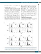
Figure 4. CD9 and CD26 cell expression among CXCR4 cell subpopulations. (A) CD9 expression at day 0 and after 7 days of culture. (B) CD26 expression at day 0 and after 7 days of culture. The same CD34+CXCR4neg, CD34+CXCR4low and CD34+CXCR4bright subpopulations were selected as those for the in vivo reconstitution experiments. Percentages of CD9+ or CD26+ cells are indicated for each subpopulation. Day 7: cell subpopulations were defined among the progeny of total day-0 CD34+ cells.
haematologica | 2018; 103(10)
1609
