
Page 42 - 2018_10-Haematologica-web
P. 42
V. Lapostolle et al.
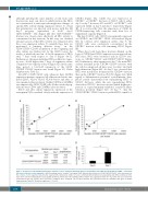
although injecting the same number of cells from each fraction into mice can show in which fraction the SRCs are concentrated, it cannot provide insight into changes of specific SRC activity during expansion culture. To obtain this information, we injected each mouse with the full day-7 progeny (equivalent) of 2x10⁵ day-0 CD34+CXCR4low cells (Figure 2D) (the CD34+CXCR4low fraction was chosen since effectively all SRC activity is concentrated in this fraction). In this way, we obtained unequivocal proof that SRC activity was enhanced after the ex vivo expansion culture. To quantify these data, we performed a limiting dilution assay31 on the CD34+CXCRlowCD133+ population at the beginning and after culture (see further text for the CD133 issue), the results of which showed an ~4.2-fold expansion of SRCs after 7 days with respect to day 0 (Figure 3A-C). Furthermore, the mean individual SRC proliferative capac- ity was ~4-fold higher after 7 days of expansion culture compared to the capacity at day 0 (Figure 3D). In the same time period, a 14.2-fold expansion of the CD34+ CXCR4lowCD133+ fraction was found (Figure 3C; Online Supplementary Table S2).
All (100%) SS-PB CD34+ cells, whatever their CXCR4 expression pattern, expressed all adhesion molecules ana- lyzed (LFA-1, VLA-4, VLA-5, VLA-6) before and after ex vivo expansion (data not shown). Neither CD90 nor CD45RA was expressed by CD34+ SS-PB cells: all the sorted subpop- ulations were CD90- and CD45RA- (data not shown).
Before and after culture expansion, expression of the tetraspanin CD9 correlated closely with the expression of
CXCR4 (Figure 4A). CD26 was not expressed on CXCR4neg or CXCR4low fractions of CD34+ cells at either day 0 or day 7, however 22% and 38% of CXCR4bright cells expressed CD26 on day 0 and day 7, respectively (Figure 4B). It is noteworthy that the expression of CD26 by CXCR4-expressing cells coincides with their loss of engraftment capacity (Figure 2).
On day-0 CD34+ cells, CD133 was primarily expressed on CXCR4neg and CXCR4low CD34+ cell fractions (Figure 5A). In contrast, after expansion culture (day 7), the CD133+ cells were exclusively concentrated in the CXCR4low fraction of the cells remaining CD34+ (Figure 5B).
When day-0 cells from these fractions defined on the basis of CXCR4 and CD133 expression were injected into NSG mice, SRCs were evidenced only in CD133+ frac- tions, i.e. CXCR4negCD133+ and CXCR4lowCD133+ (Figure 6A). Furthermore, after expansion at day 7, the main SRC activity remained in the CXCR4lowCD133+ fraction: with the cell dose employed, all mice were “positive” and with high chimerism (Figure 6B). At day 0, we observed that SRCs were much more frequent in the CXCR4low fraction than in the CXCR4neg fraction (P<0.01) (Figure 4A). With regard to differentiation potential, a predominant “lym- phoid” profile characterized the repopulating HSCs of CD133+ fractions, while the rare repopulating HSCs detected in CD133neg fractions showed much higher pro- portion of, or predominantly exhibited, a myeloid differ- entiation potential (Figure 6A). At day 7, only the CD34+CXCR4lowCD133+ fraction yielded HSCs capable of
AB
CD
Figure 3. Frequencies and individual proliferative capacity of severe combined immunodeficiency repopulating cells within the CD34+CD133+CXCR4low cell popula- tion before and after ex vivo expansion. (A,B) Percentage ofmice “positive” for human CD45, 8 weeks after injection of CD34+CXCR4lowCD133+ cells, with respect to the cell dose, before expansion (A) and after expansion (B). (C) Absolute number of severe combined immunodeficiency repopulating cells (SRCs) estimated on the basis of the extreme limiting dilution assay (ELDA). (D) Mean chimerisms of the individual SRCs (only the doses giving less than 37% of positive mice were taken into consideration and only positive mice from these conditions were analyzed). The results presented were generated from the individual data given in Online Supplementary Table S2. Statistical significance: *P<0.05.
1608
haematologica | 2018; 103(10)
