
Page 41 - 2018_10-Haematologica-web
P. 41
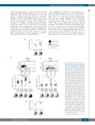
Steady-state blood CD34+ HSCs are CXCR4lowCD133+
and the end stage of the ex vivo culture (day 7) (~67% cells expressing CXCR4) (Online Supplementary Table S1). In all experiments, three distinct subpopulations of cells with respect to CXCR4 expression level were evidenced: CXCR4neg, CXCR4low and CXCR4bright (Online Supplementary Figure S1). Flow cytometry analysis after sorting showed that the cells belonged to only one of the subpopulations, categorized according to CXCR4 expression (Online Supplementary Figure S2). Most cells with in vivo repopulat- ing capacity in the day-0 population were predominantly concentrated in the CXCR4low fraction, although some minor activity was found in the CXCR4neg and CXCR4bright populations (Figure 2B); these HSCs exhibit a lower lym- phoid differentiation potential compared to CXCR4neg and, especially, CXCR4low repopulating HSCs.
The engraftment of CXCR4neg cells prompted us to explore the hypothesis that at least some of the CXCR4neg cells can express CXCR4 once in an in vivo microenviron- ment of 37°C (i.e. after injection and transplantation). Thus, after an overnight incubation, 30% of the CD34+ cells that were initially CXCR4neg, became CXCR4low (Online Supplementary Figure S3). These data from the bulk CD34+ cultures were confirmed in the cultures initiated with the sorted CXCR4neg cells (Online Supplementary Figure S4). After ex vivo expansion, almost all cells with engraft- ment capacity (SRCs) were concentrated in the CXCR4low fraction (Figure 2C) and fully maintained their day-0 dif- ferentiation potential, although from these results it appears that SRC activity after ex vivo expansion (day-7) is lower than that of non-expanded cells at day 0. However,
B
C
A
D
Figure 2. CXCR4 expression is related to the engraftment capacity of hematopoietic stem cells contained in the steady-state peripheral blood CD34+ cell population before and after ex vivo expansion. (A) The activity of severe combined immunodefi- ciency repopulating cells (SRCs) in CD34+ steady-state peripheral blood (SS-PB) cells is enhanced by 7 days of culture. Day 0: 2x10⁵ SS-PB CD34+ cells were injected per mouse. Day 7: the total progeny of 2x10⁵ day-0 SS-PB CD34+ cells was injected per mouse. (B-C) After culture day-0 (B) and day-7 (C) culture, three cell subpopulations were selected for injection into the recipient mice: P1, cells defined and sorted as the CD34+CXCR4neg subpopulation; P2, cells defined and sorted as the CD34+CXCR4low subpopulation; P3, cells defined and sorted as the CD34+CXCR4bright subpopulation. Day 0: 1x10⁵ SS-PB total CD34+ cells or 1x10⁵ cells from each sorted cell subpopulation were injected per mouse. Day 7: 2x10⁵ of the total expanded cell population or 2x10⁵ cells from each sorted cell subpopulation were injected per mouse. (D) Effect of ex vivo expansion on SRC activity in SS-PB CD34+CXCR4low cells: Day 0: 1x10⁵ SS-PB CD34+CXCR4low cells were injected per mouse; day 7: the total progeny of 1x10⁵ day-0 SS-PB CD34+CXCR4low cells were injected per mouse. (A-D) SRC activity was evaluated by short-term reconstitution (8 weeks) in NSG mice; each point represents the percentage of CD45+ human cells in one mouse bone marrow. For each condi- tion (A-D) the “pie” graphs show the relative proportion of CD19+ and CD33+ cells of human origin within the huCD45+ popula- tion. Statistical significance: *P<0.05; **P<0.01; ***P<0.001.
haematologica | 2018; 103(10)
1607
