
Page 40 - 2018_10-Haematologica-web
P. 40
V. Lapostolle et al.
positive results due to control isotype, we used the non-injected mice to establish the “positivity threshold” for CD45, which was 0.1%.24,27 Furthermore, to avoid inhibition of CXCR4 activity due to fixation of clone 12G5 antibody on the same external loop as SDF- 1α (CXCL12, CXCR4 specific ligand),28 we performed antibody elu- tion by acid solution. For this, cells were incubated 20 min at 0°C in ACDA, pH 5 (Anticoagulant Citrate Dextrose solution formula A, Bioluz, Saint-Jean-de-Luz, France), then washed twice in RPMI medium before injection into mice. All cell suspensions were treat- ed in an identical manner before injection.
To determine the SRC frequency, limiting dilution analysis29,30 was performed for the CD34+CXCR4lowCD133+ subpopulation. The details are given in the Online Supplementary Methods.
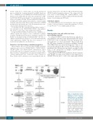
ii) Long-term HSCs (LT-HSCs). For the detection of LT-HSCs, sec- ondary recipient mice (Figure 1) were conditioned as primary recipients. The BM from both femora of primary recipients was flushed, resuspended and injected intrafemorally6,25 into the sec- ondary recipient NSG mice (Figure 1), as described in detail in the Online Supplementary Methods. The mice were sacrificed 7 or 8 weeks later and analyzed as described above.
Detection of colony-forming committed progenitors
Thawed CD34+ cells were selected for their CXCR4 and CD133 expression and sorted as CXCR4negCD133-, CXCR4negCD133+, CXCR4lowCD133- and CXCR4lowCD133+ subpopulations. Day-0 sorted subfractions were expanded separately ex vivo for 7 days. Day-0 and day-7 subpopulations were plated in methylcellulose cytokine-supplemented kits “Stemα-1D” (Saint Clement les Places, France) (1000 cells/mL for each cell population) and cul-
tured for 14 days (37°C, 20% O2, 5% CO2) in 35 mm Petri dishes (NUNC, Roskilde, Denmark) in duplicate. The colonies (>50 cells) were scored19 as burst-forming unit - erythroid (BFU-E), colony- forming unit - granulocyte and macrophage (CFU-GM) and multi- lineage colony-forming unit (CFU-mix).
Statistical analysis
The Mann-Whitney test for non-parametric values was applied. P values <0.05 were defined as statistically significant (*). P<0.01 (**) and P<0.001 (***) were highly significant values.
Results
Hematopoietic stem cells with short-term reconstituting capacity
To estimate ST-HSC activity directly before and after ex vivo expansion, the mice were injected with 2x10⁵ day-0 SS-PB CD34+ cells or with their total day-7 progeny, here- after referred to as “day-0 equivalent”. These results from the NSG mice confirmed our previous findings obtained with NOD/SCID mice6 demonstrating that 7 days of cul- ture greatly enhanced SRC activity (P<0.05) while it also maintained the differentiation potential, as judged on the basis of the proportion of lympho (CD19)-myeloid (CD33) chimerism (Figure 2A).
Regarding the SS-PB CD34+ population, the most promi- nent changes in culture were related to the expression of CXCR4 between day 0 (~16% cells expressing CXCR4)
Figure 1. Experimental design. Evaluation of human stem cells by employing a severe combined immunodeficiency repopulating cell assay before (day 0) and after (day 7) ex vivo expansion culture. (A) Human cell chimerism in pri- mary mice recipients reflects the activity of short-term hematopoiet- ic stem cells (ST-HSC) while (B) human cell chimerism in second- ary mice recipients reflects the activity of long-term hematopoietic stem cells (LT-HSC). HPC: hematopoietic progenitor cell; PC: precursor cell; MC: mature cell.
1606
haematologica | 2018; 103(10)
