
Page 45 - 2018_10-Haematologica-web
P. 45
Steady-state blood CD34+ HSCs are CXCR4lowCD133+
CD133- fractions before and after ex vivo expansion con- tained exclusively erythroid progenitors (BFU-E) irrespec- tive of CXCR4 expression, while CD133+ cells always contained three classes of progenitors (CFU-GM, BFU-E and CFU-Mix). Furthermore, the committed progenitors were five times less concentrated in the CXCR4lowCD133- fraction than in the CXCR4lowCD133+ one.
Discussion
The findings presented in this article clearly show that ST-HSCs and LT-HSCs present in SS-PB have particular
phenotypic properties, which are different from those of HSCs in CB, BM or mobilized peripheral blood (M-PB). It is evident that the pattern of CXCR4 expression is related to the functional abilities of SS-PB ST-HSCs and LT-HSCs. This is not surprising since CXCR4 and its ligand SDF-1 have been demonstrated to have a major role in hom- ing/mobilization of HPCs and HSCs.33,34
The presence of a small fraction (8%) of CXCR4- expressing CD34+ cells in SS-PB was first observed by Lataillade et al.35 Here, we found 16% of CXCR4+CD34+ cells in the mononuclear SS-PB fraction issued from leukodepletion filters. Only a very small fraction of M-PB CD34+ cells express CXCR4; these cells exhibit an in vitro
AB
C
D
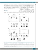
Figure 7. Capacity of long-term hematopoietic reconstitution of NSG mice is restricted to the CD34+CXCR4lowCD133+ steady-state peripheral blood cell fraction. Before (day 0) (A, C) and after ex vivo expansion (day 7) (B, D), three cell populations were selected and sorted: total CD34+ steady-state peripheral blood (SS-PB) cells, the CD34+CXCR4lowCD133+ selected subpopulation, and total CD34+ cells without the CD34+CXCR4lowCD133+ subpopulation. (A) Day-0, short-term reconstitution in primary recipients, 2x10⁵ cells of each subpopulation were injected intravenously per mouse. (B) Day-7, short-term reconstitution in primary recipients, 1.6x10⁶ cells of each sub- population were injected intravenously per mouse. Day-7 subpopulations were defined among the progeny of total day-0 CD34+ cells. (C) Day-0, long-term reconstitution in secondary recipients. Bone marrow cells from both femora of each primary recipient were injected into the bone marrow of the secondary recipient NSG mouse. (D) Day-7, long-term reconstitution in secondary recipients. Bone marrow cells from both femora of each primary recipient were injected into the bone marrow of the sec- ondary recipient NSG mouse. For each condition (A-D) the “pie” graphs show the relative proportions of CD19+ and CD33+ cells of human origin within the huCD45+ pop- ulation. Statistical significance: *P<0.05; **P<0.01; ***P<0.001.
haematologica | 2018; 103(10)
1611
