
Page 46 - 2018_10-Haematologica-web
P. 46
V. Lapostolle et al.
AB
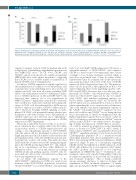
Figure 8. Hematopoietic committed progenitors in the CD34+ cell populations selected on day 0 on the basis of CXCR4 and CD133 expression. Day-0 (A) sorted sub- populations were expanded separately ex vivo and day-7 (B) clonogenic capacities of these subpopulations were analyzed. CFU-GM: colony-forming unit - granulocyte/monocyte; BFU-E: burst-forming unit – erythroid; CFU-mix: colony-forming unit - mixed. Statistical significance: *P<0.05; **P<0.01; ***P<0.001.
capacity to migrate towards a SDF-1α gradient and result in high levels of multilineage engraftment upon injection into NOD/SCID mice.10 In CB, both CXCR4+ and CXCR4neg subsets were shown to be capable of engrafting NOD/SCID mice with similar frequencies36 suggesting that CXCR4 is not a suitable marker for purification of human CB HSCs before transplantation.
In addition, CXCR4 expression is rapidly regulated by environmental factors or induced ex vivo by cytokines such as granulocyte colony-stimulating factor (also used in our cultures) in CD34+ cells from all sources, including SS-PB CD34+ cells with primitive features.35 Furthermore, induc- tion of CXCR4 expression on CB and M-PB CD34+ cells increased their capacity for in vivo engraftment in NOD/SCID mice.37 Apparently contradictory conclusions were reached in a study that found that the homing effi- ciency of CD34+ cells selected from BM or M-PB was not related to expression of either CXCR4 or adhesion mole- cules.38 However, blocking CXCR4 signaling on trans- planted CB CD34+ cells prevented homing, whereas pre- treatment of cells with cytokines led to up-regulation of CXCR4 expression and increased mice engraftment,39 which clearly highlights the crucial importance of CXCR4 expression for HSC engraftment.
This point is important because it helps to avoid erro- neous conclusions due to artifacts induced by the techni- cal procedure related to CXCR4 expression-based cell sorting; a low repopulating capacity of CD34+CXCR4+ cells (the authors did not discriminate between low and bright populations) from CB and BM could result from the neutralizing activity of the anti-CXCR4 monoclonal anti- body that was used for cell sorting. This antibody (clone 12G5) binds the site that serves for the binding and signal- ing of the CXCR4 specific ligand SDF-1α28,39 and, depend- ing on its concentration, can have either inhibitory or stimulatory effects.11 To avoid any artifacts in assaying CXCR4 activity and grafting capacity of CD34+ cells, we used antibody elution after cell subfraction sorting and before any engraftment assays.
In our hands, the engraftment capacity of the CD34+CXCR4neg subfraction in SS-PB was much lower than that of the CD34+CXCR4low population, as found for CB CD34+CXCR4neg versus CD34+CXCR4pos cells (including
both “low” and “high” CXCR4 expression).12 However, a substantial number of CD34+CXCR4neg cells expresses CXCR4 (i.e. they become “low expressing”) after at least overnight ex vivo cytokine treatment, an effect which is even more pronounced after 4 days in culture (Online Supplementary Figure S3; confirmed also in the cultures ini- tiated with the CD34+ cells sorted on the basis of CXCR4 expression: Online Supplementary Figure S4). This phenom- enon could be proposed as the explanation for the ex vivo culture enhancing effect on the engrafting capacity of ST- HSCs and LT-HSCs. However, this is not the case, since practically all CXCR4neg cells which became CXCR4+ in culture lost their CD133 expression by day 4 of culture (only CD133+ cells exhibited engrafting capacity, see below) (Online Supplementary Figure S3). However, the CD133 expression was maintained for at least for 24 h in culture mimicking the in vivo situation after cell injection. This could explain some minor engraftment capacity of the CXCRneg cell population before expansion. In fact, these cells could become CXCR4low in vivo, during the first hours after injection. It should be emphasized that the highest engraftment capacity before (at day 0) or after (at day 7) cell culture is concentrated in the CXCR4low fraction and not in the CXCR4bright one, which seems to be surpris- ing. Actually, the expression of CD26 (that, in our proto- col, could be induced by granulocyte colony-stimulating factor, as shown for CD34+CD38- CB cells40), which is related only to CXCR4bright cells, can explain the decrease in CXCR4bright engrafting efficiency. It has been shown that CD26/dipeptidyl peptidase IV is a membrane-bound extracellular peptidase that cleaves polypeptides such as SDF-1, thus reducing CXCR4 activity. Furthermore, CD26 expression might be part of a mechanism regulating CXCR4 activity. The inhibition of CD26 expression on CB CD34+ cells enhances the in vitro migratory effect against the SDF-1 gradient9 and improves in vivo long-term engraftment in NOD/SCID mice.41,42 Furthermore, pre- treatment of mice with a specific CD26 inhibitor (diprotin A) enhances engraftment of mouse BM cells in primary and secondary recipients.43 This is being considered among emerging strategies to improve homing and engraftment of HSCs in clinical transplants.44 A similar approach, allowing the CXCR4bright HSCs to engraft, might
1612
haematologica | 2018; 103(10)
