
Page 57 - 2018_09-Mondo
P. 57
MΦ-mediated BM failure
Aberrant podoplanin expression during SAA drives HSC loss, thrombocytopenia, and death
Macrophages negatively regulated both HSCs and Mks, and we questioned whether Mfs also regulate non- hematopoietic BM stromal cells, known to be impaired in SAA patients.30-32 In contrast to radiation alone, SAA reduced osteoblastic and endothelial cells (Online Supplementary Figure S7A and B). Because SAA was associ- ated with severe thrombocytopenia we examined expres- sion of podoplanin (PDPN), recently identified in the BM and shown to increase platelet production.33 We noted a striking loss in PDPN+ stromal cells in SAA (Online Supplementary Figure S7C and D). At the same time, how- ever, we observed induction of PDPN on hematopoietic cells that appeared to be entirely restricted to Mfs and a majority were CD11blo/- Mfs (Figure 6A and B, and Online Supplementary Figure S8A). We found no change in PDPN expression among other hematopoietic or non- hematopoietic stromal cells (data not shown). In addition, we observed increased podoplanin (gp38) transcripts specifically in the CD11blo/- Mfs, relative to CD11b+ Mfs, T cells, and neutrophils in SAA mice (Figure 6C). PDPN+ Mfs were reduced in MIIG mice relative to controls, though PDPN MFI was unchanged (Online Supplementary
AB
Figure S8B and C). This suggests that IFNγ increased num- bers of PDPN+BM Mfs, rather than directly regulating PDPN expression, during SAA.
To determine if aberrant PDPN expression on Mfs mediated pathology during SAA, we administered an anti- PDPN monoclonal antibody during SAA. PDPN blockade significantly increased CD41lo/int and CD41hi HSCs and resulted in a preservation of BM cellularity compared to isotype control treatment (Figure 7A and B). Administration of an anti-PDPN antibody did not rescue HSCs by reducing or impairing T-cell activation because similar numbers of T-bet+ CD4 and CD8 T cells and IFNγ levels were observed in the BM of anti-PDPN and control- treated mice during SAA (Online Supplementary Figure S9A and B). Consistent with improved HSC numbers PDPN blockade rescued thrombocytopenia, increased BM Mks, and increased survival in SAA (Figure 7C-E). Anti-PDPN antibody conferred significant protection, whereas iso- type-control antibody-treated mice had a median survival of only 16.5 days and died between days 12 and 19 induc- tion of SAA.
PDPN-CLEC-2 interaction was reported to induce RANTES,33 which can support platelet production, thus our finding that PDPN blockade improved thrombocy-
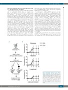
Figure 5. Macrophage depletion increases platelet- biased hematopoietic stem cells (HSC) in severe aplastic anemia (SAA). (A) HSC function in SAA was assessed by transplantation of HSCs from PBS- (TM) or clod-lip (Clod;˜)-treated TdTomato+ F1 mice 8 days post-splenocyte transfer (d.p.s.t.). (B) Peripheral blood was analyzed for reconstitution at indicated time points. P<0.0001 for platelets, P=0.002 for myeloid, P=0.06 for lymphoid. Each transplantation data set represents one experiment, n=4-5 recipient mice per group. Two-way ANOVA was used to compare between groups.
haematologica | 2018; 103(9)
1457
