
Page 58 - 2018_09-Mondo
P. 58
A. McCabe et al.
AB
C
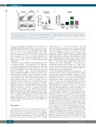
Figure 6. Macrophages exhibit increased expression of podoplanin (PDPN) during severe aplastic anemia (SAA). (A) PDPN expression in bone marrow (BM) cells ++ ++
(top) 8 days post-splenocyte transfer (d.p.s.t.). CD11b expression among PDPN F4/80 cells (bottom). (B) PDPN F4/80 MΦ numbers in healthy (●), radiation con- trol (■), and SAA (+Rad +Splenocytes; □) mice 8 d.p.s.t. (C) Gp38 expression in sort-purified BM populations, relative to β-actin and normalized to expression in neutrophils. Data represent data pooled from 3 independent experiments n=5-10 mice/group.
topenia was somewhat surprising. Consistent with a role for PDPN in RANTES production we observed increased RANTES during SAA (Online Supplementary Figure S9C), where we also observe increased PDPN+ Mfs. PDPN blockade did not impact BM RANTES in SAA, likely because the antibody clone (8.1.1) does not interfere with CLEC-2 binding in vivo or in vitro (Online Supplementary Figure S9D-F).34 Thus, PDPN-dependent HSC loss and hematopoietic failure occurs via a RANTES-independent mechanism.
Administration of anti-PDPN antibody induced a specif- ic decrease in CD11blo/- Mfs whereas CD11b+ Mf num- bers were not significantly different (Figure 7F). This sug- gests PDPN signaling may be important for CD11blo/- Mf survival during SAA. It also demonstrates that selective reduction of CD11blo/- Mfs is associated with improved survival during SAA. PDPN can bind and activate ezrin, radixin, and moesin family proteins to promote cytoskele- tal reorganization and contractility of fibroblastic reticular cells in lymph nodes.34 Microenvironmental stiffness can reduce physical support for HSCs and Mks,35 and we observed reduced expression of a-smooth muscle actin (aSMA), a marker of contractile stress fibers,36 by PDPN+ BM Mfs at day 8 p.s.t. upon anti-PDPN treatment (Online Supplementary Figure S10A and B). We also noted a striking increase in expression of arginase-1, a marker of M2-polar- ized Mfs, in both CD11blo/- and CD11b+ Mfs upon anti- PDPN treatment (Online Supplementary Figure S10C). Thus, CD11blo/- Mfs aberrantly express PDPN in the BM during SAA, correlating with hematopoietic failure. Future stud- ies are warranted to determine the precise impact of PDPN-expressing Mfs on the microenvironment and whether stiffness and Mf-activation state contribute to SAA pathology.
Discussion
Hematopoietic stem cell loss and BM destruction are key features of SAA, and are associated with cytokine pro- duction by T cells.6-8 It is still unclear, however, if inflam- mation depletes HSCs directly or does so through the microenvironment. Findings from SAA patient BM sug- gest that stromal support of hematopoietic cells is signifi-
cantly reduced.30-32,37,38 In a mouse model of SAA, we observed reduced stromal cells, but, at the same time, BM Mfs were maintained. CD11blo Mfs exhibited a unique survival advantage in SAA that correlated with their expression of PDPN. Consistent with our findings, SAA patient BM exhibited CD169+ Mfs persistence despite significant reductions in nearly all other hematopoietic cell types.31 Our findings reveal that, rather than direct IFNγ- mediated HSC depletion, IFNγ signaling in Mfs promotes HSC loss during SAA. IFNγ and Mfs limit CD41hi HSCs during disease, thus contributing to severe thrombocy- topenia and mortality in SAA (Figure 7G). To the best of our knowledge, this is the first in vivo study addressing the mechanistic role of the BM microenvironment in HSC loss and disease progression during SAA.
HSCs reportedly undergo apoptosis during SAA,39 yet studies in models of infection suggest that excessive differ- entiation and reduced self-renewal contribute to IFNγ- dependent HSC depletion.40,41 We previously identified an IFNγ-dependent increase in monopoiesis during Ehrlichia muris infection, which occurred at the expense of HSCs.42- 44 Monocytes are increased early in SAA, prior to their ulti- mate loss, supporting the idea that increased IFNγ-driven HSC differentiation contributes to HSC loss. It is also pos- sible that increased apoptosis in SAA is a product of enhanced differentiating divisions that render HSCs more susceptible to inflammatory stress and/or cell death.
Aberrant immune cell function, specifically T-cell activa- tion and homing to the BM, is associated with SAA.7 Since IFNγ primes Mfs for activation,25 and Mfs produce cytokines and present antigen to T cells, we predicted that IFNγ signaling in Mfs increase T-cell activation. SAA pro- gression is mitigated in MIIG mice, however, despite sim- ilar numbers of activated and IFNγ-secreting donor T cells in the BM. Cytokines associated with SAA (TNFa, IL-1β, and IFNγ) were also similarly induced. Thus, resident Mfs do not appear to drive disease through their capacity to present antigen to T cells or general inflammatory dispo- sition. Mf polarization can contribute to disease through exaggerated inflammation and wound healing responses.28 During SAA, differential expression of M1-associated Nos2 was observed between the MIIG model and anti- PDPN treatment and between CD11b+ and CD11blo/- Mfs, indicating functional differences between these two Mf
1458
haematologica | 2018; 103(9)
