
Page 55 - 2018_09-Mondo
P. 55
MΦ-mediated BM failure
Clodronate-liposomes specifically deplete macrophages, increase CD41hi HSCs and platelets, and rescue survival during SAA
models, a similar and significant increase in megakary- ocyte progenitors was observed in MIIG SAA mice and Mf-depleted SAA mice (Online Supplementary Figure S4B).
A similar rate of platelet removal from circulation was observed in PBS- and clod-lip-treated SAA mice (Figure 4H and I). Thus, improved platelet counts were not due to loss of consumption by Mfs. The increase in BM Mks and significantly reduced mortality in Mf-depleted mice com- pared to PBS-lip-treated controls demonstrate Mfs drive SAA mortality, possibly via their ability to restrict pheno- typically-defined platelet-biased HSCs. Thus, HSC loss and thrombocytopenia is dependent on Mfs and Mf growth factors in SAA.
T-cell responses are not impaired in clod-lip-treated and MIIG mice
Macrophages may drive HSC loss by enhancing activat- ed T-cell infiltration into the BM; therefore, we tracked donor T cells by inducing SAA with splenocytes from UBC-GFP mice (C57BL/6 background). Expression of the T-helper 1 transcription factor T-bet, which is expressed in T cells of SAA patients and increases IFNγ gene transcrip- tion,7 was not diminished in T cells from Mf-depleted mice (Online Supplementary Figure S5A). IFNγ protein levels (Online Supplementary Figure S5B) and IFNγ-secreting donor T cells (Online Supplementary Figure S5C-E) in the BM of SAA mice were also unaffected by Mf depletion. T-
To test the impact of Mf depletion on SAA pathogene- sis, we administered clodronate-encapsulated liposomes (clod-lip) to mice one day after SAA induction. BM Mfs were significantly and specifically reduced 8 d.p.s.t. (Figure 4A). Monocytes and neutrophils are also phagocyt- ic and may be transiently depleted; however, they were quickly replaced and no sustained depletion was observed with clod-lip. Mf depletion correlated with improved cel- lularity at day 15 (Figure 4B and C), increased total HSCs (Online Supplementary Figure S3A), and increased CD41hi HSCs at 8 and 15 p.s.t (Figure 4D), thus supporting the idea that Mfs negatively regulate HSCs during SAA. Macrophage-colony stimulating factor (M-CSF) is critical for tissue Mf survival and self-renewal,22,23 and similar to clod-lip administration, M-CSFR antagonism significantly increased HSCs during SAA (Online Supplementary Figure S3B). Similar to MIIG SAA mice, CD41hi HSCs correlated with increased circulating platelets, significantly increased BM Mks, and improved survival (Figure 4E-G). HSCs were increased in both MIIG SAA and Mf-depleted SAA mice, while more downstream progenitors, including short- term HSCs and multipotent progenitors (MPPs), were more variable (Online Supplementary Figure S4A). Consistent with improved thrombocytopenia in both
ABC
DEF
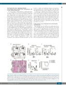
Figure 3. MIIG mice exhibit increased CD41hi hematopoietic stem cells (HSC), increased megakaryocytes in the bone marrow (BM), and reduced mortality during
aplastic anemia. (A) CD41 expression on BM HSCs in radiation control (Rad) (top) and severe aplastic anemia (SAA) (bottom) MIIG and LC mice 15 days post-spleno-
cyte transfer (d.p.s.t.). CD41 mean fluorescence intensity (MFI) on HSCs is shown on the plots and gates represent CD41lo/int and CD41hi HSCs. (B and C) CD41lo/int and
CD41hi HSC numbers in MIIG (▲) versus LC (D) mice on days 8 (mean is shown) and 15 (median is shown, and a Mann-Whitney test was used to compare between
+ groups).*P<0.05,**P<0.01.(D)Gp1bβstaininginBMofRadandSAAMIIG(▲)andLC(r)mice15d.p.s.t.Scalebar=100μm.(E)GP1bβ megakaryocytesper100
mm of sternal BM. Mean±Standard Error of Mean (SEM) is shown. **P<0.01. (F) Kaplan-Meier survival curve for radiation control (Rad; LC and MIIG mice; □, n=4) and2SAA MIIG (▲; n=9) and LC (D; n=11) mice. Log-rank (Mantel-Cox) test was used to compare between groups. *P<0.05.
haematologica | 2018; 103(9)
1455
