
Page 222 - Haematologica Vol. 107 - September 2022
P. 222
LETTER TO THE EDITOR
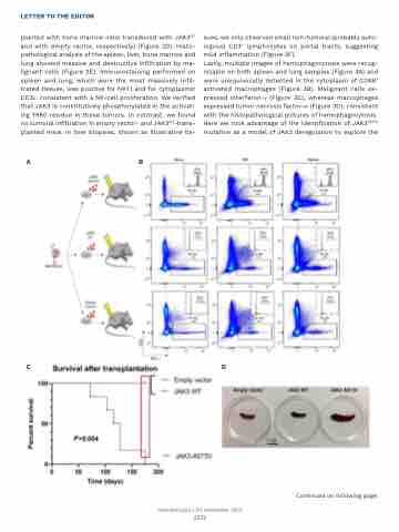
planted with bone marrow cells transduced with JAK3WT and with empty vector, respectively) (Figure 2D). Histo- pathological analysis of the spleen, liver, bone marrow and lung showed massive and destructive infiltration by ma- lignant cells (Figure 2E). Immunostaining performed on spleen and lung, which were the most massively infil- trated tissues, was positive for NK1.1 and for cytoplasmic CD3ε, consistent with a NK-cell proliferation. We verified that JAK3 is constitutively phosphorylated in the activat- ing Y980 residue in these tumors. In contrast, we found no tumoral infiltration in empty vector- and JAK3WT-trans- planted mice. In liver biopsies, shown as illustrative tis-
sues, we only observed small non-tumoral (probably auto- logous) CD3+ lymphocytes on portal tracts, suggesting mild inflammation (Figure 2F).
Lastly, multiple images of hemophagocytosis were recog- nizable on both spleen and lung samples (Figure 3A) and were unequivocally detected in the cytoplasm of CD68+ activated macrophages (Figure 3B). Malignant cells ex- pressed interferon-γ (Figure 3C), whereas macrophages expressed tumor necrosis factor-a (Figure 3D), consistent with the histopathological pictures of hemophagocytosis. Here we took advantage of the identification of JAK3A573V mutation as a model of JAK3 deregulation to explore the
AB
CD
Haematologica | 107 September 2022
2221
Continued on following page.
