
Page 224 - Haematologica Vol. 107 - September 2022
P. 224
LETTER TO THE EDITOR
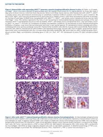
Figure 2. Natural killer cells expressing JAK3A573V generate a massive lymphoproliferative disease in mice. (A) Eight- to 10-week- old Rag2-/- donor mice were injected intraperitoneally with 150 mg/kg 5-fluorouracil (5-FU; Sigma-Aldrich, St Louis, MO, USA) 5 days prior to bone marrow collection from iliac bones, femora and tibiae. Bone marrow cells transduced with JAK3A573V, JAK3WT or empty vector were administered intravenously to sublethally irradiated C57BL/6 recipient mice (N=6 for each condition). (B) NK1.1+/CD3- eGFP+ cells from the peripheral blood, bone marrow or spleen of mice. Cells were harvested at the time of autopsy. (C) Survival of wild-type C57Bl/6 mice transplanted with JAK3A573V-, JAK3WT- and empty vector-transduced bone marrow cells from Rag2-/- mice. The red box represents the time of cell harvesting for immunophenotyping analysis and autopsy of the last diseased JAK3A573V-transplanted mouse as well as healthy JAK3WT- and empty vector-transplanted mice. Log-rank empty vector or JAK3WT vs. JAK3A573V, P=0.004. (D) Comparison of spleen size between conditions. (E) Hematoxylin & eosin staining showing the infiltration of spleen, lung, bone marrow (sternum), and liver in peri-portal spaces. (F) Hematoxylin & eosin staining and immu- nostaining with CD3ε (primary antibody: clone SP7, ThermoFisher Scientific SAS, Illkirch, France), NK1.1 (clone PK136, ThermoFisher Scientific SAS, Illkirch, France) and PY-JAK3 (D44E3, Cell Signaling Technology, Danvers, MA, USA) of spleen, lung and liver tissue. Specimens were counterstained with the corresponding secondary antibodies. BM: bone marrow; eGFP: enhanced green fluor- escent protein. Rag2: recombination activating gene 2; LSK: Lin-, Scahi, Kithi. HE: hematoxylin & eosin; PY-JAK3: phosphorylated JAK3.
Figure 3. Mice with JAK3A573V-induced lymphoproliferative disease develop hemophagocytosis. (A) Macrophages phagocytosing cells (red arrows) in spleen and lung tissue. (B) CD68 stain (clone 514H12, Leica Biosystems, Nanterre, France) shows numerous macrophages as large, irregularly shaped CD68+ cells. Detail of CD68 stain shows active phagocytosis by CD68+ macrophages of lymphocytes (red arrows). (C) Interferon-γ stain (clone ab216642, Abcam, Paris, France) shows interferon-γ production by malig- nant cells. (D) Tumor necrosis factor-a (TNF-a) stain (AF410-NA, R&D System, Minneapolis, MN, USA) shows TNF-a production by macrophages, identified as large irregularly shaped cells with an abundant cytoplasm. A detailed morphology of TNF-a-se- creting macrophages is provided. HE: hematoxylin & eosin; IFN-γ: interferon-γ. TNF-a: tumor necrosis factor-a.
Haematologica | 107 September 2022
AB
C
D
2223
