
Page 221 - Haematologica Vol. 107 - September 2022
P. 221
LETTER TO THE EDITOR
CD
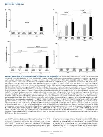
Figure 1. Generation of murine natural killer cells from LSK progenitors. (A) Experimental procedure. Five 6- to 10-week-old C57BL/6H mice were pooled in each experiment. Freshly isolated bone marrow cells were treated with Fc-block (CD16⁄CD32) and stained with biotin-conjugated lineage antibodies (CD3 [145-2C11], Gr-1 [RB6-8C5], B220 [B-220], and TER-119 [TER-119]). Lineage-positive cells were first depleted by magnetic-activated cell separation using Streptavidin Microbeads (BD Biosciences, Le Pont de Claix, France). Cells were then stained with anti-CD3-APC/Cy7, anti-Gr1-APC/Cy7, anti-B220-APC/Cy7, anti-TER119- APC/Cy7, anti-Sca1-FITC (E13-161.7), and anti-c-Kit-PerCP/Cy5.5 (2B8) and LSK cells were sorted with a Beckton Dickinson FAC- SInflux. All antibodies were purchased from Ozyme (Saint Quentin en Yvelines, France), except for biotin-conjugated lineage antibodies and the anti TER119-APC/Cy7, which were purchased from BD Biosciences (Le Pont de Claix, France). LSK cells were then transduced with JAK3A573V, JAK3WT or empty vector as previously described8,9 and cultured in 96-well plates on MS5 stromal cells for 10 days in Dulbecco modified Eagle medium with murine stem cell factor (25 ng/mL), murine thrombopoietin (10 ng/mL), human interleukin-7 (10 ng/mL), murine interleukin-15 (50 ng/mL) (all from PeproTech, Rocky Hill, NJ, USA) and human Fms-like tyrosine kinase 3-ligand (10 ng/mL) (Celldex Therapeutics, Inc., Needham, MA, USA), for natural killer (NK)-cell differentiation. Cells were then stained with anti-CD3-APC, anti-NK1.1-PerCp/Cy5.5, anti-NKp46-PECy7, as well as intracellular Alexa Fluor® 647 fluorochrome-conjugated antibodies for PY-STAT3 (Y705), PY-STAT5 (Y594) or PY-ERK 1/2 (Y202/204) or phospho-AKT (S473) after permeabilization with Perifix expose purchased from Beckman Coulter (Villepinte, France), according to the manufacturer’s in- structions. (B) The bar chart shows that the number of mature murine NK cells (CD3-, NK1.1+, NKp46+) obtained from 104 LSK cells transduced with JAK3A573V, JAK3WT or empty vector, after 10 days of culture. Results are the mean ± standard deviation of three independent experiments. The statistical significance was calculated by a Kruskal-Wallis test. (C) Analysis of intracellular staining for Y705-STAT3, Y594-STAT5, Y202/204-ERK1/2, and S573-AKT. Results are provided in percentages from CD3- NK1.1+ NKp46+ NK cells. LSK: Lin-, Scahi, Kithi; WT: wild-type; mSCF: murine stem cell factor; mIL-3: murine interleukin-3; hFlt3-L: human Fms-like tyrosine kinase 3-ligand; hIL-7: human interleukin-7; mIL-15: murine interleukin-15; hIL-2: human interleukin-2; NK: natural killer; GFP: green fluorescent protein.
or JAK3WT remained alive and disease-free (log-rank test, P=0.004) (Figure 2C). Moreover, the blood cell count of two sick JAK3A573V mice before autopsy showed pancytopenia, with the lymphopenia and thrombocytopenia being par-
ticularly pronounced (Online Supplementary Table S2), a hallmark of hemophagocytic syndrome.10 Autopsy of these two mice was remarkable for the spleen enlargement (492±285 mg vs. 86±9 mg and 82±9 mg in mice trans-
Haematologica | 107 September 2022
2220
