
Page 92 - 2022_03-Haematologica-web
P. 92
J.T. Weinreb et al.
Ddx41 deficiency triggers cell cycle arrest in erythroid progenitors
The diminished number of erythroid progenitors and dysregulated expression of cell cycle genes suggest that defects in erythrocyte proliferation could contribute to
the anemia in ddx41 mutants. In order to examine prolif- eration, we analyzed cell cycle status of 30 hpf gata1:dsred+ erythroid progenitors by flow cytometry quantification of DNA synthesis via EdU incorporation and DNA content via DAPI incorporation. The ddx41
AB
CD
E
FGH
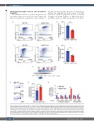
Figure 2. Ddx41 regulates erythroid progenitor numbers. (A and C) Flow cytometry plots of gata1:dsred+ erythroid cells from sibling controls (left) and ddx41 mutants (right) at 28 days post fertilization (dpf) (A) and 40 hpf (C). (B and D) Graphs depicting the absolute number of gata1:dsred+ erythroid cells per embryo from (A) and (C), respectively. n=5 pools of ~5-20 embryos per pool. (E) Schema of erythroid-myeloid progenitor (EMP) development. ProE: proerythroblasts; BasoE: basophilic ery- throblasts; OrthoE: orthochromatophilic erythroblasts; func. Ery: functional erythrocytes. (F) In situ hybridization of cmyb at 26 hpf in sibling controls (left) and ddx41 mutants (right) (scale bars=150 μm). (G) Quantification of cmyb PBI in situ hybridization levels from (F). Quantification was done using Fiji; n=10-12 embryos. Graphs display means ± standard deviations (stds). (H) Graph of reverse transcription quantitative polymerase chain reaction (RT-qPCR) analysis of the expression of globin genes between sibling controls and ddx41 mutants. Expression levels were normalized to slc4a1 levels. Graph displays means ± standard error mean. The P-values were calculated with an unpaired t-test, *P<0.05, **P≤0.01, ***P≤0.001; n=3 replicates per genotype.
648
haematologica | 2022; 107(3)
