
Page 90 - 2022_03-Haematologica-web
P. 90
J.T. Weinreb et al.
A
BCDE
FG
H
J
I
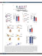
Figure 1. Loss of ddx41 causes anemia. (A) Schema of primitive erythroid development, PLM: posterior lateral mesoderm; ProE: proerythroblasts; BasoE: basophilic ery- throblasts; OrthoE: orthochromatophilic erythroblasts; func. Ery: functional erythrocytes. (B and D) In situ hybridization of the erythroid markers cmyb (B) (scale bars =200 μm) and gata1 (D) (scale bars =250 mm] at 22 hours post fertilization (hpf) in sibling controls (top) and ddx41 mutants (bottom). Arrowheads highlight the intermediate cell mass (ICM) region in the embryos. (C and E) Quantification of c-myb (C) and gata1 (E) in situ hybridization levels from (B) and (D), respectively. Quantification was done using Fiji. (F and H) Staining for o-dianisidine, marking functional hemoglobin in mature primitive erythrocytes, in sibling controls (left) and ddx41 mutants (right) at 40 hpf (F) (scale bars =350 mm) and 48 hpf (H) (scale bars =400 mm). Numbers on bottom left corner indicate the fraction of embryos with the same phenotype as the one depicted in the image. (G) Graph depicting size of erythrocytes in sibling controls and ddx41 mutants at 40 hpf. (I) Graph depicting frequency of designated o- dianisidine staining levels in sibling controls and ddx41 mutants at 48 hpf. (J) Representative images of orthochromatophilic erythroblasts stained with May–Grunwald– Giemsa from sibling controls (left) and ddx41 mutants (right) at 48 hpf (scale bars =5 mm). Graphs display means ± standard deviations (stds) with P-values calculated with unpaired Student’s t-test, ns=not significant (P>0.05), ****P>0.0001. For in situs and o-dianisidine staining n=6-72 embryos per experiment.
646
haematologica | 2022; 107(3)
