
Page 54 - 2022_03-Haematologica-web
P. 54
M. Zapatka et al.
A
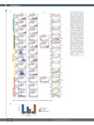
Figure 4. Changes in cancer cell fractions and evolution types. (A) Evolution patterns in 12 patients. Probability dis- tribution of the cancer cell fraction (CCF) for each somat- ic single nucleotide variant (SNV) revealed clonal (red) or subclonal (blue) SNV (left side). The changes in CCF are depicted in the last column. Changes (a mutation with a change in CCF of greater than 0.2 (DCCF>0.2) with probabil- ity >0.5) are highlighted in green (increased CCF) or red (reduced CCF). On the basis of time dependent changes of CCF (right side), evolution pat- terns were considered as (unbranched) co-evolution (C) when significant changes were unidirectional (up or down), or branched (B) when significant changes were in both directions (up and down) indicating that a dominant clone is replaced by its sib- lings. Time between sam- plings is indicated in years at the top. (B) Difference in occurrence of evolution types across clinical phases (co- evolution = blue, branched evolution = brown).
B
610
haematologica | 2022; 107(3)
