
Page 103 - 2022_03-Haematologica-web
P. 103
Dendritic cells in myelodysplastic syndromes
Figure 1. Cell subset enumeration in myelodysplastic syndrome- and healthy-derived bone marrow and peripheral blood. (A) Gating strategy of dendritic cells (DC) and slan+ non-classical monocytes in normal bone marrow (NBM) and myelodysplastic syndromes (MDS)-derived BM. After debris and doublet exclusion, CD45+ mononuclear cells were gated. Then plasmacytoid DC (pDC), myeloid DC (cDC1 and cDC2) and slan+ monocytes were identified based on the expression of CD141high, CD1c and M-DC8/CD16, respectively. (B) Frequencies of different cell subsets in normal bone marrow (NBM) compared to MDS BM. In total 30 NBM samples and 187 MDS BM samples were used. Percentages were calculated from the mononuclear cell fraction. Mean frequencies ± standard error of the mean (SEM) are given (NBM vs. MDS BM: pDC 0.76% SEM ± 0.09 vs. 0.91% SEM ± 0.11, cDC1 0.048% SEM ± 0.006 vs. 0.030% SEM ± 0.003, cDC2 0.67% SEM ± 0.05 vs. 0.54% SEM ± 0.04 and slan+ 0.36% SEM ± 0.07 vs. 0.24% SEM ± 0.02). (C) Cell frequencies in different classification groups according to the 2016 World Health Organization (WHO) classification. Patients having a higher blast count-related 2016 WHO classification (EB-1/EB-2) show lower percentages of DC and slan+ monocytes compared to NBM and lower risk groups (SLD/MLD/RS-SLD/RS-MLD). NBM (n=30) vs. (RS-)SLD/MLD (n=115) vs. EB-1/EB-2 (n=48): pDC 0.76% vs. 1.11% vs. 0.63%, cDC1 0.048% vs. 0.038% vs. 0.015%, cDC2 0.67% vs. 0.59% vs. 0.44%, slan+ 0.36% vs. 0.24% vs. 0.23%. (D) Cell frequencies in different risk groups within the International Prognostic Scoring System (IPSS). The percentages of myeloid DC subsets decrease gradually in higher risk groups. NBM (n=30) vs. low risk (n=49) vs. intermediate-1 (n=71) vs. intermediate-2 (n=21) vs. high risk (n=5): cDC1 0.048% vs. 0.035% vs. 0.031% vs. 0.010% vs. 0.005%, cDC2 0.67% vs. 0.57% vs. 0.52% vs. 0.42% vs. 0.26%. (E) Cell frequencies in different risk groups within the IPSS-R. Again, differences between sub- groups are mainly seen in DC subsets. Higher risk groups show lower percentages of DC compared to NBM and lower risk groups. NBM (n=30) vs. very low/low risk (n=77) vs. intermediate risk (n=32) vs. high/very high risk (n=27): cDC1 0.048% vs. 0.038% vs. 0.019% vs. 0.013%, cDC2 0.67% vs. 0.62% vs. 0.41% vs. 0.35%. (F) Correlation of cell frequencies in MDS-derived peripheral blood (PB) and BM samples. In total, 26 paired MDS samples were included. The non-para- metric Spearman’s correlation test was used to find significant correlations between frequencies in PB and BM. *P<0.05, **P<0.01, ***P<0.001, ****P<0.0001. EB: excess blasts; MLD: multilineage dysplasia; RS-MLD: ring sideroblasts with multilineage dysplasia; RS-SLD: ring sideroblasts with single lin- eage dysplasia; SLD: single lineage dysplasia.
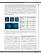
Figure 2. Clonal involvement of dendritic cells subsets and slan+ monocytes. Fluorescence in situ hybridization (FISH) analysis of sorted cells, including B cells and CD34+ blast cells, with a known cytogenetic aberrancy. In three tested cases (monosomy 7, del 5q and trisomy 8), isolated cDC2 and CD34+ blast cells were highly involved in the dysplastic clone, whereas B cells were not involved. Slan+ monocytes showed clonal involvement in del5q. Interphase FISH on whole bone marrow samples showed both an aberrant and a normal cell line. A representative FISH analysis is shown in which interphase cells are hybridized with the chromosome 5q probe displayed in red and 5p probe displayed in green (LSI EGR1(5q31)/D5S23,D5S721(5p15.2) Dual Colour Probe Set). Loss of 5q is seen in CD34+ blasts, cDC2 and slan+ monocytes (2G1R), but not in B cells (2G2R).
complement receptors (C3AR1) are at the top of this list. Further leading-edge analysis using all five gene sets was performed to identify the genes that highly account for the gene set’s enrichment signal (Figure 4A). There was a great overlap of genes that formed the leading-edge sub- set between all gene sets (Figure 4B). In total, 418 genes were found to form the leading-edge subset for cDC2 of which 32 were present in all five gene sets. For slan+ monocytes 353 genes formed the leading-edge subset. Of them, 39 genes were found in all five gene sets. These genes were considered most relevant because they form the core of the enrichment (Figure 4C). Again, both lists with lead targets consisted of multiple pattern recogni- tion receptors, which suggests an overall diminished capacity for sensing pathogen/damage associated molec- ular patterns (PAMP/DAMP) by MDS-derived cells. This was further confirmed by the fact that also genes that were highly involved in subsequent down-stream cell signaling, such as BTK, CARD9, IRAK4, IRF3/7, MyD88, SYK, and usually lead to activation of pro-inflammatory processes, were down-regulated in MDS-derived APC.
Myelodysplastic syndrome-derived cells show reduced T-cell priming capacities and clear Th1/2-type T cell skewing
Next, in order to confirm the hypothesis that was formed from the gene expression profiling data, functional capacities of cDC2 and slan+ monocytes were tested. Upon stimulation with LPS and R848, a combination of proven synergistically working TLR ligands,33 cDC2 showed upregulation of co-stimulatory molecules. In con- trast, slan+ monocytes were unable to upregulate matura- tion markers (Figure 5A). Again, cDC1 were not tested because of low frequencies. There was no statistical differ- ence in maturation capacity for cDC2 when they were compared to NBM-derived cDC2. Slan+ monocytes showed a significantly reduced ability to upregulate CD80 upon stimulation compared to their equivalents in NBM (Figure 5B). In order to investigate their cytokine secreting capacity, MDS-derived cDC2 and slan+ monocytes were isolated and either left non-stimulated or stimulated overnight with TLR ligands. Culture supernatants were tested for the presence of different cytokines. Compared
haematologica | 2022; 107(3)
659
