
Page 89 - 2022_02-Haematologica-web
P. 89
iNKT cells promote tolerance by cDC apoptosis
A
B
C
D
E
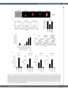
Figure 3. Culture-expanded invariant natural killer T cells require cell contact to induce apoptosis through degranulated effector molecules. (A) Representative image stream assay illustrating direct cellular contact between invariant natural killer T (iNKT) cells (PBS57-loaded CD1d tetramer+, yellow) and dendrtic cells (DC) (HLA-DR+, pink) and subsequent DC apoptosis induction (annexin V+, green) after 4 hours (h) of co-incubation. (B) Representative dot plots showing DC apoptosis and pooled data of living DC (annexin V-propidium iodide [PI]-) after co-incubation with iNKT cells either directly or separated by a transwell insert (TW). (C) Percentage of DC apoptosis inhibition after blocking of the receptors CD1d, FasL, TRAIL, NKG2D and applying the inhibitors zVAD-fmk (N-benzyloxycarbonyl-Val-Ala-Asp(O-Me) flu- oromethylketone), CMA (concanamycin A) and monensin/brefeldin A. (D) Representative dot plots showing DC apoptosis after co-culture with non-degranulated and degranulated iNKT-cell supernatant. (E) IFN-γ, granzyme B, perforin and granulysin release by iNKT cells after encountering DC analyzed by bead-based immunoas- say. Histograms show the mean of three independent experiments (n=3). Error bars show standard error of the mean. ns: not significant; *P<0.05, **P<0.01, ***P<0.001; HLA-DR: human leukocyte antigen DR-isotype.
haematologica | 2022; 107(2)
431
