
Page 87 - 2022_02-Haematologica-web
P. 87
iNKT cells promote tolerance by cDC apoptosis
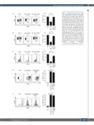
Figure 1. Culture-expanded invariant natural A killer T cells inhibit T-cell activation and prolifer- ation. Representative dot plots and histograms showing (A) early activated T cells (CD69+, day 1), (B) late activated T cells (CD25+, day 3) and (C) proliferating T cells (carboxyfluorescein suc- cinimidyl ester [CFSE], day 7). T-cell activation and proliferation was measured after incubation with monocyte-derived dendritic cell (mo-DC) in the presence or absence of invariant natural killer T (iNKT) cells. iNKT cells were added to the culture either directly or separately through a transwell insert (TW). (D) Representative dot plots and histograms showing late activated T cells (CD25+, day 3) and (E) proliferating T cells (CFSE, day 7) after stimulation with anti- CD3/CD28-coated beads in the presence or absence of iNKT cells. All events were gated on single cells and living lymphocytes. iNKT cells were excluded from the analysis by gating on CD3+ PBS57-loaded CD1d tetramer+ popula- tions. Histograms show the mean of three inde- pendent experiments (n=3). Error bars indicate standard error of the mean. ns: not significant, *P<0.05, **P<0.01, ***P<0.001,
B
C
D
****P<0.0001. DC: dendritic cells; T: T cells.
E
haematologica | 2022; 107(2)
429
