
Page 206 - 2022_02-Haematologica-web
P. 206
Letters to the Editor
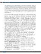
Figure 3. Continuous exposure to permethrin or chlorpyrifos during pregnancy fails to induce Mll breaks in bone marrow progenitors or alterations in the hematopoietic homeostasis of mothers, pups or adult offspring in mice. (A) Experimental design to determine whether prenatal exposure to etoposide (ETO), permethrin (PER) or chlorpyrifos (CPF) induces Mll breaks or hematopoietic alterations in mothers, pups or adult offspring in mice. In brief, pregnant CD1 female mice were exposed to PER (20 mg/kg/day), CPF (20 mg/kg/day), ETO (10mg/kg/day) or 0.1 % dimethylsulfoxide (DMSO) from day 0 to day 21 of gestation. The number of pups per litter and their sex distribution were analyzed at birth. The mothers and one-half of the litter were analyzed at weaning while the remaining one-half of the offspring were maintained for 32 weeks for analysis in adulthood. (B) Upper panel, scheme depicting mouse chromosome 9 and the region where mouse Mll probes hybridize (UCSC, GRCm38/mm10). Lower left panel, a representative fluorescence in situ hybridization (FISH) image of a mouse metaphase and presence of fluorescence signals in both Mll alleles. Lower right panel, a zoom image of mouse chromosome 9 by DAPI banding, with and without the BAC fluorescence signal revealing the Mll gene localization. (C) Percentage of mouse bone marrow (BM) LK cells with Mll breaks detected by interphase FISH at sac- rifice. DMSO and ETO were used as negative and positive controls, respectively. The numbers in bars indicate the number of mice analyzed. (D) Fluorescence activated cells sorting (FACS) BM analysis of the Lin- Sca-1+ Kit+ (LSK) subpopulation (upper panels), hematopoietic stem and progenitor cell subsets (middle panels), and mature cells (lower panels) in mothers, pups and adult offspring. (E) Representative macroscopic images of spleens (upper panel) and livers (lower panel) at sacrifice of mothers, pups and adult offspring exposed to the indicated treatments. HSC: hematopoietic stem cells; MPP: multipotent progenitors; HPC: hematopoietic progenitor cells.
ences in the proportions of mature cells (myeloid, T and B) in peripheral blood or total Lin-Sca+Kit+ progenitors, hematopoietic stem cells, multipotent progenitors or hematopoietic progenitor cells in the bone marrow between treatments in any group (Figure 3D, Online Supplementary Figure S3D). Similarly, prenatal exposure to etoposide or pesticides did not affect the hematopoi- etic homeostasis in peripheral blood, as determined by absolute numbers of white blood cells, red blood cells and platelets (Online Supplementary Figure S3E). Lastly, no evidence of splenomegaly or hepatomegaly was observed in mothers, pups or adult offspring (Figure 3E). Our results thus suggest that chronic exposure to perme- thrin or chlorpyrifos during pregnancy does not induce Mll breaks in bone marrow progenitors or alterations in the hematopoietic homeostasis of mothers, pups or adult offspring.
A unique strength of the present study is the cutting- edge in vivo models employed to assess the genotoxicity and leukemogenesis potential of etoposide, permethrin or chlorpyrifos. The NSG mice model was established to mimic the adult exposure associated with occupational risk, whereas the CD1 mice model attempted to mimic prenatal exposure to topoisomerase II poisons and insec- ticides suggested to be involved in the etiology of infant leukemia. Continuous exposure to permethrin or chlor- pyrifos in both models failed to induce MLL breaks or alterations in hematopoietic homeostasis, confirming the in vitro results of limited genotoxicity and no leuke- mogenic potential of permethrin or chlorpyrifos in human and murine HSPC after chronic exposure. The fact that MLL breaks are acutely induced by permethrin or chlorpyrifos but are not sustained upon long-term chronic exposure in vitro or in vivo indicates a legitimate repair of the DNA damage/DSB in the MLL locus. Of note, although long-term in vivo exposure to etoposide did induce MLLr in some hematopoietic progenitors, it failed to initiate leukemia in either in vivo models, in line with a previous study confirming that in utero exposure to etoposide did not trigger the development of leukemia in either Atm+/+ or Atm-/- mice.13 The eventual develop- ment of overt leukemia might depend on the survival and proliferative advantage of minor MLLr pre-leukemic clones, targeting the right cell-of-origin, on stromal bone marrow interactions and also on the acquisition of sec- ondary cooperating oncogenic alterations. The clearance and lack of selection of MLLr clones is consistent with the development of MLLr treatment-related acute leukemia in adults or infant leukemia in only a rare subset of patients exposed to topoisomerase II poisons.
Our results clearly suggest that permethrin and chlor- pyrifos induce MLL breaks in human HSPC across ontogeny. However, such insecticide-induced DNA-DSB are successfully repaired, and do not involve chromoso-
mal translocations encoding MLL fusions. This explains their limited genotoxicity and no leukemogenic potential and reinforces why these compounds are still considered as non-classifiable carcinogens. Linking environmental or genotoxic exposure to causal and/or functional MLL chromosomal translocations has long been controversial,11 and the cutting-edge in vitro and in vivo cellular models employed in the present study also have obvious limitations. For instance, site-specific cleavage within the MLL break cluster region (bcr) has been shown to be induced by either topoisomerase II poisons but also genotoxic chemotherapeutic agents which do not target topoisomerase II and even by non-genotoxic stimuli of apoptotic cell death. In addition, MLL chromo- somal translocations have been linked to higher-order chromatin fragmentation that occurs during the initial stages of apoptosis, suggesting that the generation of MLL chromosomal translocations (and likely others) are part of a generalized acute apoptotic response-mediated higher-order chromatin fragmentation which ultimately renders a chromosome topology and chromatin struc- ture prone to chromosomal DNA exchanges.14 This is further supported by the ambiguity of MLL transloca- tions partnering with a large number of different chro- mosomal loci.
Virginia C. Rodriguez-Cortez,1 María Pilar Navarrete- Meneses,2* Oscar Molina,1* Talia Velasco-Hernandez,1 Jessica Gonzalez,3 Paola Romecin,1 Francisco Gutierrez- Agüera,1 Heleia Roca-Ho,1 Meritxell Vinyoles,1 Eric Kowarz,4 Pedro Marin,5 Sandra Rodriguez-Perales,6 Carlos Gomez-Marin,7 Patricia Perez-Vera,2 Felipe Cortes- Ledesma,7 Anna Bigas,1,3,8 Andrea Terron,9 Clara Bueno1,8 and Pablo Menendez1,8,10
1Josep Carreras Leukemia Research Institute. Department of Biomedicine. School of Medicine, University of Barcelona. Barcelona, Spain; 2Laboratorio de Genética y Cáncer, Departamento de Genética Humana, Instituto Nacional de Pediatría, Ciudad de México, México; 3Cancer Research Program, Institut Hospital del Mar d'Investigacions
4 Mèdiques, Hospital del Mar, Barcelona, Spain; Institute of
Pharmaceutical Biology/DCAL, Goethe-University of Frankfurt, Frankfurt/Main, Germany; 5Hematology Department. Hospital Clínic de Barcelona, Barcelona, Spain; 6Molecular Cytogenetics Group, Human Cancer Genetics Program, Spanish National Cancer Research Center (CNIO), Madrid, Spain; 7Topology and DNA Breaks Group, Spanish National Cancer Center (CNIO), Madrid, Spain; 8Centro de Investigación Biomedica en Red-Oncología (CIBERONC), Madrid, Spain; 9European Food and Safety Authority. Parma. Italy and 10Instituciò Catalana de Recerca i Estudis Avançats (ICREA), Barcelona, Spain.
*MPNM and OM contributed equally.
Correspondence:
PABLO MENÉNDEZ - pmenendez@carrerasresearch.org
548
haematologica | 2022; 107(2)
