
Page 270 - 2022_01-Haematologica-web
P. 270
M. Faleschini et al.
co-transfected in Meg01, as previously described.2 The luciferase activity was detected using the Dual Luciferase reporter assay sys- tem kit (Promega), according to the manufacturer’s instructions. Details of the experiments are included in the Online Supplementary Appendix.
Quantitative real-time polymerase chain reaction
Quantitative real-time polymerase chain reaction (qRT-PCR) was performed to evaluate the absolute expression of GFI1B iso- forms and relative expression of GFI1B major target, using specific primers (Online Supplementary Table S2). FastStart Universal SYBR Green Master Mix (Roche) and ABI PRISM 7900 detection (Applied Biosystem, Foster City, CA, USA) were used. All experi- ments were performed three times in triplicate, as previously reported.22
Western blot analyses
HEK293T cells were transfected with myc-tagged wild-type and mutant GFI1B cDNA using calcium phosphate method and main- tained in DMEM medium (Euroclone, Pero, Italy) with 10% fetal bovine serum. Protein fractionated cell extracts were analyzed by western blot using primary antibodies anti-myc (9E10; sc-40; Santa Cruz Biotechnology, Dallas, TX, USA), anti-HSP90 (sc-7947; Santa Cruz Biotechnology), anti-ORC2 (ab68348; Abcam, Cambridge, UK); as previously described.23
Immunofluorescence assay
GFI1B transfected in HeLa cells was detected using 9E10 anti- body against c-myc (Santa Cruz Biotechnology). Peripheral blood smears were double-labeled with antibodies against CD41 (Santa Cruz Biotechnology) and CD34 (Miltenyi Biotec, Bergisch Gladbach, Germany). General staining and image acquisition have previously been described.24,25 Detailed methods for both immuno- fluorescence analyses are included in the Online Supplementary Appendix.
Results
Identification of the novel c.648+5G>A mutation in
GFI1B
WES of proband II-2 revealed a single nucleotide het- erozygous substitution (c.648+5 G>A) in the GFI1B gene, affecting the splice donor site of intron 9 (Figure 1A). Sanger sequencing confirmed the variant in the proband (II-2), her sister (II-3) and her daughters (III-1 and III-2) but not in her
healthy sister (II-5) (Figure 1B). The proband’s brother (II-4) was homozygous for the wild-type allele and the thrombo- cytopenia was likely due to liver cirrhosis. The c.648+5G>A substitution is reported in GnomAD with a microcytic anemia factor (MAF) value of 0.000008025 and identified in two European (non-Finnish) individuals in het- erozygous status.
Based on NNSplice software, the c.648+5G>A variant is predicted to drop the score of the splice donor site from 0.89 to 0.24, suggesting potential alternative splicing processes. Therefore, RT-PCR analysis was performed using RNA extracted from affected individuals’ peripheral blood cells (II-2 and III-1) (Figure 1C). In contrast with the healthy control showing the expected product of 246 base- pairs (bp), the patients’ samples amplified an additional band of 108 bp. Sequencing analysis revealed that the lower band corresponded to a transcript characterized by skipping of exon 9, resulting in an in-frame mRNA deletion of 138 bp (p.Val171_Gln216del). The alternative splicing is expected to produce a shorter protein of 32kDa, lacking zinc fingers 1 and 2, corresponding to what was previously described as p32.7,26
Blood cell studies in family members
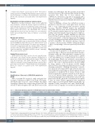
The major blood parameters of the family members are reported in Table 1. The propositus had moderate thrombo- cytopenia while the other individuals carrying the het- erozygous c.648+5G>A GFI1B mutation had a platelet count only slightly lower or higher than the lower limit of the normal range. Platelet size calculated as mean platelet volume (MPV) by the automated counter was normal in all subjects. The study of myeloproliferative disorders (MPD) by image analysis of blood smears revealed slightly increased values, indicating mild platelet macrocytosis. The remaining parameters, including hemoglobin concentra- tion, mean red cell volume (MCV) and red blood cell count were within the normal ranges in all investigated subjects. Examination of peripheral blood smears revealed a mild reduction in platelet a-granules and red blood cell anisocy- tosis in all affected family members. Flow cytometry revealed normal expression of the platelet surface GP com- plexes Ia-IIa, IIb-IIIa, and Ib-IX-V in individuals II-2 and III- 1 (data not shown). Platelet aggregation after stimulation with collagen (4 and 20 mg/mL), ADP (5 and 20 mM,) epi- nephrine (10 mM), arachidonic acid (1 mM), TRAP (25 mM), and ristocetin (1.5 mg/mL) was normal in individuals II-2
Table 1. Clinical and laboratory parameters in GFI1B mutated and non-mutated family members
Subject (Sex)
II-2 (F)
II-3 (F)
III-1 (F)
III-2 (F)
II-4 (M)
II-5 (F) Normal values
GFI1B Platelet mutational count
status (x109/L)
648+5G>A/+ 50 648+5G>A/+ 149 648+5G>A/+ 124 648+5G>A/+ 153
+/+ 94* +/+ 215
150-450
MPV (fL)
10.7 9.2 12.6 11.7 10.8 10 8-13**
MPD (mm) 3.32
2.95 2.96 3.57 3.09 3.26 2.4-2.7†
Hb (g/dL)
13.6
13.4
12
13.9
15.6
14.6
11.7-15.5 (F) 13.2-17.3 (M)
MCV (fL)
85 90.6 82.3 88.3 98 95.1 82-98
RBC (x106/mL)
4.89
4.40
4.10
4.80
4.92
4.58
3.8-5.2 (F) 4.4–5.7 (M)
RCDW (%)
14 13.7 13.4 15 13.9 14 11.6-16
Leukocyte WHO count bleeding
(x109/L) score§
6.23 3 4.01 3 7.62 0 9.1 0 5.7 0 5.42 0 4-10 0
£F: female; M: male; * in this patient thrombocytopenia was likely due to liver cirrhosis; **MPV range given by the automated cell counter; †MPD obtained in 50 healthy volunteers (95% confidence interval)18; §World Health Organization (WHO) bleeding scale: 0 - no bleeding tendency; 1 - cutaneous bleeding only (including minimal mucosal bleeding); 2 - mild blood loss (any mucosal bleeding not fulfilling the criteria for grade 1 or 3); 3 - gross blood loss, requiring transfusion; 4 - debilitating blood loss (including retinal or cerebral associated with fatality). See patients and methods for details about bleeding symptoms. MPV: mean platelet volume; MDP: myeloproliferative disorders; Hb: hemoglobin; RBC: red blood cell.
262
haematologica | 2022; 107(1)
