
Page 269 - 2022_01-Haematologica-web
P. 269
AB
C
Novel GFI1B variant dysregulates oncogenic factors
ulation of the respective gene at the transcriptional level.10 Since its first description,2 individuals with GFI1B muta- tions have been examined in order to characterize this novel rare platelet disorder. To our knowledge, these stud- ies have identified at least 15 different mutations in approx- imately 20 unrelated families, allowing better characteriza- tion of the disease, whose features are increased bleeding tendency, thrombocytopenia with enlarged platelets and a reduced a-granule content with abnormal distribution.2,3,10–17 We report a novel heterozygous GFI1B variant (c.648+5G>A) in a family with mild thrombocytopenia and no other significant features. The substitution causes skip- ping of exon 9 and consequent overexpression of the short p32 isoform and dysregulation of GFI1B target genes, including CD34 and other genes that are involved in neo- plastic transformation, suggesting a potential role for GFI1B
in carcinogenesis regulation.
Methods
Family study and clinical features
The propositus (Figure 1), a 65-year-old female with low platelet number, and her family members were studied to determine the molecular basis of thrombocytopenia. Their clinical features, as well as the methods used for blood cell analyses,18,19 are described in more detail in the Online Supplementary Appendix. The Institutional Review Board of the IRCCS “Policlinico San Matteo
Foundation” of Pavia approved the study. All subjects provided written informed consent for the study, which was conducted in accordance with the Declaration of Helsinki.
Mutation screening and reverse transcriptase polymerase chain reaction analysis
Mutational screening was performed by whole exome sequenc- ing (WES) in the proband (II-2), as previously reported.20 Variants were confirmed by Sanger sequencing in her daughters (III-1 and III-2), sisters (II-3 and II-4) and brother (II5) (Table 1). Total RNA was extracted from peripheral blood cells of patients II-2 and III-1, as well as two healthy controls and cDNA amplified as previously reported,21 using primers enlisted in the Online SupplementaryTable S1.
Bioinformatic analysis
The effect of the splice-site mutation was predicted by in sil- ico analyses using two dedicated bioinformatic tools: splice site prediction by Neural Network (NNSplice; http://www.fruitfly.org/seq_tools/splice.html) and Human Splicing Finder Version 2.4.1 (http://www.umd.be/HSF/).
Expression vectors and dual-luciferase reporter assay
Wild-type and mutant (c.511_648del due to skipping of exon 9) GFI1B cDNA were cloned into the tagged (myc) expression vector pcDNA3.1 (Invitrogen, Carlsbad, CA, USA). The GFI1B, GFI, MEIS1, and CD34 promoters were cloned into a reporter firefly luciferase vector (pGL4/luc2, Promega). Plasmids were transiently
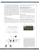
Figure 1 Genetic analyses of the family. (A) Sanger sequencing showing exon 9 (e9) and intron 9 (i9) boundary of GFI1B gene. The heterozygous c.648+5G>A substitution is indicated by an arrow. Nucleotide A of the ATG translation initiation start site of the GFI1B cDNA in GenBank sequence NM_004188.6 is indicated as nucleotide +1. (B) Real-time polymerase chain reaction showing different transcription pattern between patients (II-2; III-1) and healthy control (HC). The expected product of 246 basepairs (bp) was detected in the HC sample; an additional band of 108 bp corresponding to skipping of exon 9 was found in the affected individuals. M: molecular weight. (C) Family pedigree showing an autoso- mal dominant pattern of inheritance. Arrow indicates the proband whereas black filled symbols represent affected individuals carrying the mutation. The grey filled symbol indi- cates a thrombocytopenic brother without the mutation (platelet count in this patient may be influenced by a liver cir- rhosis). wt: wild-type.
haematologica | 2022; 107(1)
261
