
Page 233 - 2022_01-Haematologica-web
P. 233
Baseline SUVmax in FL and tumor proliferation
tures in biopsies. However, no significant correlation emerged between immune escape/T-cell activation scores and baseline SUVmax or PFS (Online Supplementary Figure S2). In order to further assess the patterns of leukocyte infil- trates in the tumor samples, we performed algorithmic deconvolution by CIBERSORT to infer the proportion of 14 leukocyte and other non-hematopoietic cell types from each sample. At the same time, we assessed the immune cell composition of FL samples by IHC. Deconvolution of
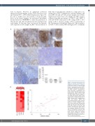
A
bulk tumor transcriptomes unveiled a composition con- taining more CD8+ T lymphocytes and macrophages and fewer NK cells and gd T-cell cytolytic lymphocytes (Online Supplementary Figure S3A). Accordingly, the IHC analyses evidenced median percentages of CD3+T cells, CD8+ T cells and CD163+ monocytes of 35% (range, 5-60%), 14% (range, 1-40%), and 11% (range, 1-30%), respectively, of the total immune cell infiltrate (Figure 4A). The abundance of CD8+ T cells scored by IHC was high (>30% of immune
B
Figure 3. Immunohistochemical vali- dation of immune escape gene set (IEGS33) overexpression in follicular lymphoma samples. (A) Upper panel shows a representative case of PD-1 staining with diffuse (left) intrafollicu- lar (right) patterns (magnification: 100x and 50x, respectively and inserts: 200x). Medium panel shows representative cases of PD-L1 (left) and LAG3 (right) staining (magnifica- tion: 100x and inserts: 200x). Lower panel shows a representative case of TIM3 staining (left) (magnification: 100x and inserts: 200x) and immunohistochemical (IHC) quantifi- cation of immune checkpoint (ICP): PD-1, PD-L1, LAG3 and TIM3 staining (right). (B) The heat map (left) repre- sents IHC scoring of the four ICP markers and the graph (right) shows the correlation between the percent- age of ICP-positive immune cells scored by IHC and the sample enrich- ment scores (SES) for immune escape gene sets (IEGS) in each FL sample. Each sample is shown by a dot. FL: follicular lymphoma.
haematologica | 2022; 107(1)
225
