
Page 64 - 2021_12-Haematologica-web
P. 64
C. Sargas et al.
(both CEBPA alleles mutated). Two variants with similar VAF were reported as probably biallelic variants (Online Supplementary Table S6). Although CEBPA variants were dis- tributed along the entire coding sequence, biallelic variants were frequently detected in BZIP (43.33%) and the N-ter- minal domain (30%). Monoallelic variants, were mostly detected in BZIP (44.83%) and almost equally distributed among TAD1 (13.79%), TAD2 (17.94%) and the N-termi- nal domain (6.90%).
IDH1 and IDH2 mutations
IDH variants were detected in 22.60% of samples. In
mutated IDH1 samples (9.11%), all variants were detected in the Arg132 codon. IDH2 (13.85%) was exclusively mutated in the Arg140 or Arg172 codons (84.20% and 15.79%, respectively). Three patients (1.61%) showed simultaneous variants in IDH1 and IDH2 (Online Supplementary Table S7). All IDH1 and IDH2 variants were targetable mutations by IDH inhibitors as no atypical vari- ants were detected.
ASXL1, RUNX1 and TP53 mutations
Overall, 39.00% of samples showed variants in at least
one of them: 18.23% RUNX1, 14.70% TP53 and 12.39% ASXL1. In 26.12% of samples a variant detected in one of these genes was the only clinically relevant variant. Moreover, 28.19% of patients were classified to an unfavor- able risk group according to ASXL1, RUNX1 and TP53 mutations, following European LeukemiaNet 2017 recom- mendations.
Discussion
This study shows that a network platform involving many highly skilled laboratories can successfully deliver robust molecular data for AML patients. This strategy allows for testing NGS in the majority of newly diagnosed and relapsed/refractory AML patients involved in the PETHEMA studies, overcoming the current challenging needs for a high-standard diagnosis in cooperative groups. Our descriptive analysis performed in a large series of real- life patients depicts the complex molecular landscape of AML.
In the last 5 years, NGS has irrupted as a potential routine tool for molecular diagnosis, allowing for precise and simul- taneous detection of relevant variants in AML. However, this technique is still non-affordable for many institutions due to: i) remarkable cost as compared to conventional PCR tests, ii) batch of samples, ranging from eight to more than 30, to run the test, and consequently high time consump- tion, both making it difficult to report results in less than 7- 10 days; and iii) the need of expensive machinery and high- ly-qualified teams for biostatistical and molecular biolo- gists. Moreover, the majority of prognostic or druggable mutations can be rapidly and relatively easily detected by conventional PCR. In fact, from the mandatory NGS panel genes selected by the PETHEMA central laboratories (i.e, ASXL1, CEBPA, FLT3, IDH1, IDH2, NPM1, RUNX1, and TP53), a mutation screening by conventional PCR is still required for FLT3, IDH1, IDH2 and NPM1, as a positive result could allow for rapid implementation of targeted or risk-adapted therapeutic approaches.8 In addition, rapid PCR is also needed for core-binding factor (CBF), PML- RARA and BCR-ABL rearrangements. Under this scenario,
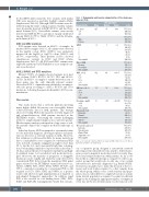
Table 3. Demographic and baseline characteristics of the study popu- lation (n=751).
NGS population
Characteristic Mean Median
Age, years 62.5 65 <60
≥60
Sex Male
Female
Range
8-93
n (%)
751 (100) 284 (38) 467 (62)
751 (100) 423 (56) 328 (44)
284 (38)
751 (100) 378 (50) 155 (21) 218 (29)
751 (100) 205 (27) 58 (8) 146 (19) 95 (13) 247 (33)
751 (100) 109 (15) 203 (27) 157 (21) 282 (38)
751 (100) 400 (53) 68 (9) 283 (38) 751 (100) 25 (3) 224 (30) 66 (9) 125 (17) 311 (41)
751 (100) 297 (40) 125 (17) 33 (4) 20 (3) 276 (37)
ECOG 0.9 1 0-4 0
1
2 48(6) 3 26(3) 4 6(1)
Not available
Type of AML
De novo
Secondary Not available
WBC, ×109/L 31.2 8.4 ≤ 5
5-10
10-50
> 50
Not available
BM blast cells, % 55 53 ≤ 30
30-70
> 70
Not available
Creatinine, mg/dL 1.0 0.87 ≤ 1,2
> 1,2
Not available Cytogenetic risk
Favorable Normal Intermediate Adverse
Not available
Therapeutic approach Intensive
Non-intensive Clinical Trial Supportive care Not available
0.3-305
2-100
0.23-3.78
751(100) 184 (25) 203 (27)
AML: acute myeloid leukemia, BM: bone marrow, WBC: white blood cell; ECOG: Eastern Cooperative Oncology Group.
our cooperative group designed a nationwide network involving seven central laboratories aimed to deliver homo- geneous and comparable molecular results for newly diag- nosed and relapsed/refractory AML patients. As far as we know, this is a different strategy as compared to other coop- erative groups that usually rely on only one or two central laboratories for molecular diagnostics (e.g, British NCRI).9 Several reasons guided us to make this decision: i) the eco- nomic and work burden required to collect samples from the whole group, which covers a wide territory and popu- lation, was not affordable for a single laboratory; ii) a mini- mum referral population is required to permit an efficient diagnosis by studying the appropriate number of samples in every run; iii) the involvement many on-site teams in order
3084
haematologica | 2021; 106(12)
