
Page 66 - 2021_12-Haematologica-web
P. 66
C. Sargas et al.
A
B
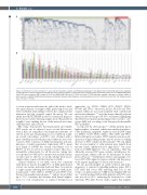
Figure 4. Distribution of gene alterations in acute myeloid leukemia samples. (A) Mutational landscape in the global cohort. Horizontal green bars: diagnosis, orange: refractory, and red: relapse.Vertical dark blue bars: positive (in global FLT3 row represents internal tandem duplications [ITD]), red: FLT3-D835/I836, green: other FLT3 point mutations (PM), orange: FLT3-ITD and D835/I836, light blue: FLT3-ITD and other FLT3-PM, light grey: negative, dark grey: not tested, yellow: bi- allelic variants in CEBPA. (B) Mutational prevalence according to disease stage. Diagnosis are represented as blue bars, refractory as red bars and relapse as green bars. *P<0.05, **P<0.01.
to create a true research network; and iv) the need to facili- tate rapid delivery of samples while preserving closer and well established relationships between the sample referral institution and the assigned central laboratory. We can affirm that the PETHEMA model for centralized diagnosis has been successful collecting samples from 751 patients in roughly 1 year, enabling the use of this network in routine clinical practice and research.
Our study demonstrates that harmonized and reliable NGS results can be achieved across several laboratories, even if they are using their own diagnostic platforms. As shown in the pre-standardization cross-validation round, an overall concordance of 60.98% was obtained with a great variability in selected genes and conditions across lab- oratories. After consensus of AML relevant genes and opti- mization of quality parameters (uniformity >85%; mean read depth of 1,000X) the overall concordance rose to 85.57% in the second cross-validation round. This was a remarkable achievement for all laboratories taking into account that low VAF (≤5%) variants were included in this second round. To the best of our knowledge, there are no similar studies reported in the literature for AML.
Clinical validation of our AML cohort was consistent with previous reports. Roughly 91% of AML patients had at least one variant, and many harbored three, four and up to eight variants reflecting the heterogeneous AML muta- tional profile.10 FLT3, IDH1/2, DNMT3A and NPM1 were the most frequently mutated genes,11,12 and we showed that up to 73% of patients had variants with clinical implica- tions for risk stratification or targeted therapy-based
approaches (i.e, ASXL1, CEBPA, FLT3, IDH1/2, NPM1, RUNX1 and TP53).13 Moreover, ASXL1, RUNX1 and TP53 variants which are not easily analyzed with conventional molecular techniques,14–16 were the unique clinically relevant alteration detected in up to 28.19% of patients, highlighting that NGS-based mutational profiling seems crucial to cate- gorize AML risk according to the European LeukemiaNet 2017 guidelines.3
As reported by other groups,17–20 elderly patients had a higher number of variants, which were enriched in spliceo- some machinery, epigenetic regulators and in DNA repair (i.e, SRSF2, U2AF1, SF3B1, ASXL1, TET2, IDH2 and TP53). In line with previous studies, NPM1 variants were more fre- quent in younger AML patients, and we noticed a striking decrease of FLT3 variants in older patients.21 We can affirm that a lower number of older patients may benefit from tyrosine kinase inhibitors-based approaches,22 but more from novel IDH-inhibitors.23 In our experience, NGS has efficiently screened FLT3 gene variants, including less fre- quent variants, which could also be informative for thera- peutic decisions.24,25 Furthermore, NGS is a promising tool to assess FLT3-ITD duplicated region, which could have prog- nostic impact regarding its location and extension.26
We also provide insights on clonal evolution and leuke- mogenesis: i) variants in signalling pathway genes (FLT3, KIT, RAS) had lower VAF, reflecting their role as late events,27; ii) genes related to CHIP showed higher VAF val- ues,28 and iii) median VAF in AML patients with TP53 vari- ants was above 50%, indicating the frequent loss of the wild-type TP53 allele. Recent studies suggest that a higher
3086
haematologica | 2021; 106(12)
