
Page 51 - 2021_12-Haematologica-web
P. 51
IL1-IL1RAP axis in AML
D
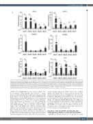
Figure 2. IL1-induced IL1RAP signaling is associated with an inflammatory secretome. (A) Transcriptome analysis of genes 2-fold upregulated in one primary acute myeloid leukemia (AML) patient (AML#1) and two AML cell lines upon stimulation with IL1b. (B) Gene ontology analysis on 331 genes that were 2-fold upregulated in at least two out of three groups (THP1, K562 and AML#1). (C) Gene set enrichment analysis analysis on a ranked gene list of AML#1. Genes were ranked from upregulated to downregulated upon stimulation with IL1b. Normalized enrichment score (NES) and false discovery rate (FDR) were used to determine significance. (D) Quantitative real-time polymerase chain reaction analysis of five primary AML patient samples ± IL1b stimulation. Bars indicate mean ± standard deviation of biological triplicates. Statistical analysis was performed by a one-tailed Student’s t-test. *P<0.05; ** P<0.01; ***P<0.001.
of JNK, p38 and MEK/ERK was less effective (Figure 3A). In accordance, K562 IL1RAP+ cells showed a strong increase in phosphorylation of p65 upon stimulation with IL1b, which could be partially reversed by inhibiting the NFkB pathway with an IKK inhibitor in a dose-dependent manner (Figure 3B to C). These data are in line with pre- vious observations by Bosman et al. who studied the TAK1-NFkB axis.17 Next, we transduced K562 IL1RAP+ cells, K562 IL1RAP– cells (as negative controls), OCI- AML3 cells, and THP1 cells with short hairpin RNA (shRNA) against IL1RAP and sorted transduced cells by green fluorescence protein (GFP) positivity (Figure 3D; Online Supplementary Figure S3E and G). Knockdown of IL1RAP did not result in impaired cell proliferation (Figure 3E; Online Supplemental Figure S3H). We observed reduced
colony-forming capacity in THP1 cells but not in OCI- AML3 cells (Figure 3F). A trend towards reduced CFC capacity upon knockdown of IL1RAP was also observed in primary AML patients #8 and #9 as also observed pre- viously (Figure 3G; Online Supplementary Figure S3I to J).28 Finally, we challenged K562, OCI-AML3 and THP1 cells by serum deprivation and stimulated them with IL1b to determine whether cell viability was controlled by the IL1-IL1RAP axis under stress conditions. Serum starva- tion-induced loss of viability, however, this was not res- cued by addition of IL1b (Figure 3H).
Synergism of the IL1-IL1RAP signaling with other active signaling pathways in acute myeloid leukemia
IL1RAP was recently described to be directly associated
haematologica | 2021; 106(12)
3071
