
Page 50 - 2021_12-Haematologica-web
P. 50
B. de Boer et al.
and IL36 depending on its co-receptor IL1R1, IL1RL1 or IL1RL2, and although, e.g., IL33 has also recently been shown to impact on HSC,26 we initially focused on IL1b in our studies. IL1b was chosen since the IL1R1 receptor appeared to be highly expressed in some AML subsets (data not shown) and due to its potential role in inflamma- tion,27 which we wanted to investigate in more detail.
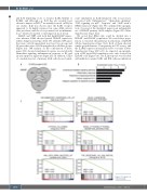
AML#1 and THP1 cells both had high IL1RAP expres- sion whereas K562 showed partial IL1RAP expression (Online Supplementary Figure S2B). We identified 299 genes that were >2-fold upregulated in at least two groups and 32 genes that were >2-fold upregulated in all three groups (Figure 2A). GO analysis on the combination of these genes (331) showed enrichment for genes associated with chemokine signaling, inflammation, response to IL1 and an anti-apoptotic signature (Figure 2B). In addition, GSEA of a ranked gene list of primary AML cells showed signif-
icant enrichment in IL1b-stimulated cells for processes associated with “inflammation”, “chemokine signaling”, “TNF signaling via p38”, “hypoxia” and “AML with a NPM1 mutation” (Figure 2C). We confirmed the upregula- tion of several of the identified genes in an independent set of IL1RAP+ primary AML samples (Figure 2D; Online Supplementary Figure S2C).
We noted that K562 cells could be divided into a IL1RAP+ and IL1RAP– population. We sorted these popu- lations to elucidate the pathways downstream of IL1RAP (Online Supplementary Figure S3A). Both of them showed similar growth kinetics, clonogenicity in CFC assays, and the IL1RAP expression remained stable over time (Online Supplementary Figure S3B to D). As expected, an upregula- tion of IL8 upon IL1b stimulation was only observed in IL1RAP+ cells (Figure 3A). The IL1b response was blocked with inhibitors against TAK1 and IKK, whereas inhibition
AB
C
Figure 2. Legend on following page.
3070
haematologica | 2021; 106(12)
