
Page 49 - 2021_12-Haematologica-web
P. 49
IL1-IL1RAP axis in AML
A
B
C
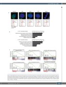
Figure 1. High IL1RAP plasma membrane protein expression in acute myeloid leukemia is associated with a leukemic granulocyte-monocyte progenitor signature.
(A) Immunofluorescent staining and expression measured by flow cytometry (red histogram) of interleukin-1 receptor accessory protein (IL1RAP) in multiple acute myeloid leukemia (AML) cell lines and one primary AML patient. The grey histogram indicates the unstained control. (B and C). Gene ontology analysis (C) and gene set enrichment analysis (GSEA) (D) on a ranked gene list based on label-free quantitative protein expression in 42 primary AML patient samples.7 Genes were ranked based on Pearson correlation with IL1RAP protein expression. Normalized enrichment score (NES) and false discovery rate (FDR) were used to determine signifi- cance. *P<0.05, ***P<0.001.
haematologica | 2021; 106(12)
3069
