
Page 52 - 2021_12-Haematologica-web
P. 52
B. de Boer et al.
AB
CDE
FG
H
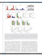
Figure 3. IL1-IL1RAP mediated activation of the NFkB signaling does not rescue proliferation under stress conditions, but IL1RAP knockdown results in reduced colony-forming capacity. (A) Quantitative real-time polymerase chain reaction (qRT-PCR) analysis in K562 IL1RAP+ and IL1RAP– cells treated with TAK1, NFkB, JNK, p38 and MEK inhibitors and subsequently stimulated with IL1b. Bars indicate mean ± standard deviation (SD) of technical triplicates. (B) Western blot of K562 IL1RAP+ and IL1RAP- treated with or without IL1b and/or IKK inhibitor (IKK inh). (C) Quantification of western blot in panel C, p-p65 was normalized to b-ACTIN. (D) Interleukin-1 receptor accessory protein (IL1RAP) mRNA levels measured by qPCR in K562 IL1RAP+ and IL1RAP- cells transduced with short hairpin (shRNA) including a non-targeting control (scr) and shRNA targeting IL1RAP (shI and shII). Bars indicate mean ± SD of technical triplicates. (E) Growth curves of K562 IL1RAP+ and IL1RAP- cells (n=3) ± knockdown of IL1RAP. (F) Colony-forming cell (CFC) output of OCI-AML3 and THP1 ± knockdown of IL1RAP. Bars indicate mean ± SD of tech- nical duplicates. (G) CFC output of acute myeloid leukemia (AML) patient 8 (AML#8) and AML#9 ± knockdown of IL1RAP. Bars indicate mean ± SD of technical dupli- cates. (H) Growth curves after serum depletion of K562, OCI-AML3 and THP1 cells (n=3) ± IL1b. Statistical analysis in all panels was performed using a Student’s t-test. *P<0.05; **P<0.01; ***P<0.001.
with FLT3 (CD135) and c-kit (CD117) receptors.28 We ana- lyzed co-expression of IL1RAP with signaling receptors including CD135, CD117 and IL3 receptor (CD123) in immature AML stem progenitor cells (CD34+ or SSClowCD117+ in case of CD34 expression <1%) by flow cytometry in a cohort of 124 primary AML patients of which four representative examples are shown (Figure 4A). Subsequently, patients were defined as single posi- tive, double positive, or double negative when at least 50% of the cells resided within either one of the gates (Figure 4A and B). These analyses revealed that 21% of the patients were CD135+IL1RAP+, while 4% expressed IL1RAP without CD135 (Figure 4B); 29.8% and 28.1% of the patients were IL1RAP+CD117+ and IL1RAP+CD123+, respectively, and we did not identify patients that
expressed IL1RAP without any detectable CD117 or CD123 (Figure 4B). Expression of IL1RAP, as quantified by flow cytometry (mean fluorescense intensity [MFI]), corre- lated significantly with CD123 expression, and to a lesser degree with CD135, but no significant correlations were found with CD117 (Figure 4C; Online Supplementary Figure 4A). These observations indicate that IL1RAP signaling can co-occur in cells that are also hardwired for FLT3 lig- and (FLT3L), stem cell factor (SCF) and/or IL3-induced sig- nal transduction, and in fact might influence those path- ways as well. In addition, Muto et al. showed that MDS HSPC switch from canonical to non-canonical NFkB sig- naling in response to inflammatory signals like IL1b, which might also occur in AML cells.29 In order to investi- gate both hypotheses, we isolated CD34+ cells from AML
3072
haematologica | 2021; 106(12)
