
Page 131 - 2021_10-Haematologica-web
P. 131
B-cell evolutionary patterns in NLPHL
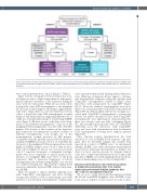
Figure 4. Schematic overview of identified malignant rearrangements in cohorts 2 and 3 with nodular lymphocyte-predominant Hodgkin lymphoma. NLPHL: nodu- lar lymphocyte-predominant Hodgkin lymphoma; DLBCL: diffuse large B-cell lymphoma; ID: initial diagnosis; Rel: relapse; Trafo: transformation; LP: lymphocyte pre- dominant.
were found in patients from cohort 3 (Figure 3, Table 1). Figure 3 shows overlap plots between repertoires at ini- tial diagnosis and at relapse/transformation, demonstrat- ing the repertoire frequency of the respective malignant clone at the two time points. While all cases from cohort 2 shared the same IGH rearrangement as the malignant LP-cell clone at initial diagnosis and relapse, suggesting the same cell of origin, only four of seven evaluable cases from cohort 3 shared the same IGH rearrangement at initial diagnosis and transformation, suggesting different cells of origin in a substantial percentage of transforming NLPHL cases (Figure 3). Of note, in two of the three cases desig- nated as “non-identical rearrangements”, only one of the malignant IGH rearrangements was detectable with FR3 primers. The absence of this sequence in the respective paired repertoire was interpreted as indirect evidence of different rearrangements even though one of the rearrangements escaped detection by FR3 primers. Compatible with the globally increased B-cell repertoire clonality in NLPHL cases from cohort 3, we found that the frequency of the malignant IGH rearrangement was much higher in NLPHL samples from cohort 3 than in those from cohort 2 (Figure 3). In four out of seven cases from cohort 3, repertoire frequencies of malignant rearrange- ments were more comparable to typical repertoire fre- quencies of a malignant NHL clone (>10% of the B-cell repertoire) than to repertoire frequencies of LP-cell rearrangements in non-transforming NLPHL cases (around 1% of the B-cell repertoire). This pointed to further B-cell repertoire similarities between NHL and pre-transforma-
tion NLPHL.
Next, we wanted to explore whether relapsing cases
with “longCDR3” rearrangements and those carrying average-sized CDR3 as well as transforming cases with identical or non-identical rearrangements at initial diag-
nosis and transformation had unifying clinical character- istics. While age at diagnosis did not appear to discrimi- nate between these respective subgroups, patients with “longCDR3” rearrangements seemed to relapse earlier than those with average-sized “no longCDR3” (Figure 5A, B). Moreover, identical rearrangements were more likely to be found in patients with earlier transformation (Figure 5B). In addition, IgD positivity was strongly asso- ciated with “longCDR3” rearrangements, as previously shown,9 but almost one-third of cases with “longCDR3” rearrangements were IgD-negative demonstrating that these two features were not fully overlapping (Figure 5C). Histomorphological patterns were also distributed differently across the subcohorts, with “longCDR3” cases and cases with transformation from an identical IGH rearrangement showing more atypical patterns (Figure5C).
Since ongoing somatic hypermutation has been described to occur in NLPHL B-cell receptors38 and anti- genic drive by Moraxella antigens has recently been reported in a subset of NLPHL cases,9 we investigated whether NLPHL cases with subsequent transformation showed more intraclonal diversification as indirect evi- dence for more selective pressure by antigens. Indeed, some level of intraclonal diversification was found in the majority of NLPHL cases with quantitatively more LP sub- clones in transforming cases of cohort 3 (Figure 5D).
Intraclonal diversification and evolutionary patterns in lymphocyte-predominatn cells from nodular lymphocyte-predominant Hodgkin lymphoma that did or did not subsequently transform
We set out to study intraclonal diversification within the LP-cell clone/NHL malignant clone (LP/NHL clone) clone in more depth in cohorts 2 and 3 in order to iden-
haematologica | 2021; 106(10)
2659
