
Page 133 - 2021_10-Haematologica-web
P. 133
B-cell evolutionary patterns in NLPHL
A
B
C
D
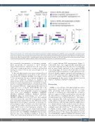
Figure 5. Characteristics of the patients with nodular lymphocyte-predominant Hodgkin lymphoma with “longCDR3” versus average CDR3 and identical versus non-identical malignant clones at initial diagnosis and relapse/transformation. (A) Age at initial diagnosis. (B) Years between initial diagnosis and relapse/transfor- mation. (C) Sex, IgD status and histological variant pattern. (D) Number of lymphocyte-predominant/non-Hodgkin lymphoma-related subclones at initial diagnosis in cohorts 2 and 3. Bars correspond to mean + standard deviation. Statistical test: unpaired, two-tailed t-test. ID: initial diagnosis; Rel: relapse/transformation; NLPHL: nodular lymphocyte-predominant Hodgkin lymphoma; DLBCL: diffuse large B-cell lymphoma; LP: lymphocyte-predominant.
tify potentially discriminative evolutionary patterns. More specifically, we intended to show whether relapse/transformation originated from the exact same cell, from a common precursor or from a cell that had undergone clonal evolution since initial diagnosis in our two cohorts.
To this end, phylogenetic trees were constructed based on somatic hypermutation exclusively for the subclones constituting the malignant LP/NHL clone (Figures 6 and 7). In some patients, the most dominant malignant sub- clone was completely identical at diagnosis and relapse/transformation (same IGH rearrangement, no dif- ference in somatic hypermutation) – eventually accompa- nied by a changing environment of clonally related, but less dominant subclones – suggesting that the same cell of origin gave rise to both the initial NLPHL and to the relapse/transformation. This pattern occurred in 11 of 18 cases, but was more frequently observed in relapsing cases (9/11 cases: NLPHL01, NLPHL03, NLPHL04, NLPHL09, NLPHL28, NLPHL13, NLPHL15, NLPHL16, NLPHL20) than in transforming cases (2/7 cases: NLPHL10, NLPHL32). In four of 18 cases, the dominant subclone(s) shared the same IGH rearrangement at diag- nosis and relapse/transformation, but differed in somatic hypermutation following a sequential evolutionary tra- jectory (Figures 6 and 7). In these cases the relapse/trans- formation clone likely developed from the same cell of origin which, however, had undergone antigenic drive between initial diagnosis and relapse/transformation. This pattern was numerically more frequent in transform- ing cases (2/7 cases: NLPHL11, NLPHL35) than in relaps- ing cases (2/11 cases: NLPHL02, NLPHL14). In three out of ten cases, transformations likely arose from a different
cell of origin (different IGH rearrangement) (Figure 7). Collectively, these data suggested that transformations more likely originated from cells that were not fully iden- tical or even clonally unrelated to the founder NLPHL clone. Intriguingly, we observed that especially cases that transformed with non-identical or fully unrelated clones showed a highly complex sequential and branching evo- lution in their initial LP clone suggesting high selective pressure by antigens which potentially extends to other rearrangements in the course of this immune reaction (Figure 7, NLPHL11, NLPHL33, NLPHL35).
Discussion
NLPHL is a rare subtype of Hodgkin lymphoma with a characteristic clinical presentation and disease course, often presenting with localized disease, favorable responses to treatment but a rather high rate of transfor- mation to aggressive NHL. Its pathogenesis and especially the characteristics of malignant LP cells as well as its B- cell environment are of increasing interest to the hemato- logic community. One of the major limitations to analysis of this entity is the paucity of LP cells in a background of normal B lineage cells that impair high-throughput genet- ic or immunogenetic studies on these clones. For this rea- son, analyses of the malignant LP cells’ BCR have been confined to microdissected cases.38-41 An interesting series of NLPHL recently caught attention since it pointed strongly to directional forces through antigenic selection in the pathogenesis of this disease. This interaction study on recombinantly expressed NLPHL BCR derived from microdissected LP cells suggested that bacterial antigens
haematologica | 2021; 106(10)
2661
