
Page 147 - 2021_09-Haematologica-web
P. 147
Functional and oncogenic roles of PRDM1 in NK cells
F
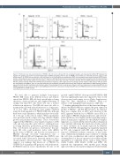
Figure 5. Proliferation and cell cycle distribution of PRDM1-/- NK cells versus wild-type NK cells. Cell growth analysis was measured by CellTiter 96® AQueous One Solution. The growth curve shows the relative fold-change of OD490 compared to the first day. (A) PRDM1 was knocked out in NK cells (donor #1) using a Cas9- sgRNA plasmid. (B) PRDM1 was knocked out of NK cells (donor #2) by Cas9/sgRNA ribonucleoprotein (RNP) and homology directed repair template (HDRT) electro- poration. Cell proliferation was analyzed with the Click-iTTM Alexa Fluor 647-EdU Flow Cytometry assay during 5 consecutive days starting from the third day after fresh irradiated feeder cells had been added. (C) Flow analysis of clones #3 and #5 on day 3 and day 7 and donor #2 cells. FITC anti-CD56 antibody was used to identify NK cells. (D) Each staining of Alexa Fluor 647-EdU was performed in duplicate, and the figure shows the average. (E) Flow analysis of EdU assay on NK cells (donor #2). GFP positivity (for PRDM1-/- cells). (F) Cell cycle distribution was analyzed by cell DNA content staining with propidium iodide/RNase or DAPI with flow cytometric assay at the third day after fresh feeder cells had been added. The percentage of cells in each phase is shown in each box. Upper panel: corresponding WT cells and PRDM1 knocked-out cells produced using the Cas9-sgRNA plasmid method. Lower panel: corresponding WT cells and PRDM1 knocked-out cells produced using the Cas9/sgRNA RNP and HDRT method.
We were able to generate multiple homozygous PRDM1 KO clones from different donors and demon- strated that PRDM1 KO cells had a much higher cloning efficiency, a faster growth rate and a higher percentage of cycling cells than WT cells. There was also a modest reduction in apoptosis. The KO cells were also able to maintain their growth and proliferation for a longer peri- od in vitro. These observations were largely validated using the bulk population of PRDM1 KO cells, although there were some differences, which may have been relat- ed to the age of the cells in culture. When experiments were performed with younger cells, they exhibited higher growth and proliferation potentials. These observations suggested that PRDM1 is a key negative regulator of NK cells, and removing this control allows a striking increase in cloning efficiency, proliferation, growth and lifespan. This corroborated our previous observations6 that pri- mary NK cells could proliferate better with shRNA knock-down of PRDM1, and that the reconstitution of PRDM1 through retroviral transduction into a PRDM1-deficient NK-cell lymphoma line (KHYG) was associated with G2/M arrest, increased apoptosis and a strong negative selection pressure. This again strongly supports our findings that knock-out of PRDM1 by CRISPR/Cas9 in primary NK promotes cell proliferation. In PRDM1 function rescue experiments, the rate of cell
growth of pMIG-PRDM1 electroporated NK PRDM1 KO cells was significantly repressed compared to that of cells electroporated with empty vector (Online Supplementary Figure S9). Thus, elimination of PRDM1 likely con- tributes to the malignant transformation of NK-cells.
To further understand the basis of the functional changes induced by PRDM1 KO, we compared the gene expression profiles of PRDM1-/- and WT NK-cell clones. As expected, there was a higher level of MYC and activation of the MYC signature with the loss of PRDM1, since MYC is a direct target of PRDM1. Many proliferation- and cell cycle- related pathways were highly enriched, including upregu- lation of many genes regulated by the DREAM complex. Signatures associated with IL-6, IL-15 and IL-2 stimulation were enriched, indicating that functional activities related to these cytokines are normally repressed by PRDM1. Similarly, TCR/NK-cell signaling was negatively regulated in WT cells compared with KO cells, indicating that removal of PRDM1 facilitates activation of these path- ways. Additional mechanisms may contribute to driving the cell cycle, proliferation and survival, including the downregulation of the pro-apoptotic factor BIM and the upregulation of additional factors such as TLR4, TOX2, CCR4, VGEFA, MYB, BCAT1, FGFR1 and SIPR1. We repeated the experiments with another gene editing approach without subsequent cloning and obtained similar
haematologica | 2021; 106(9)
2435
