
Page 145 - 2021_09-Haematologica-web
P. 145
Functional and oncogenic roles of PRDM1 in NK cells
quent presence of a large number of genetic and epigenetic changes in an established tumor as well as tumor hetero- geneity. In addition, gene expression studies of the bulk tumor contain signals from the stromal elements as well as the tumor cells. We decided to develop an approach to study selected lesions in the normal cellular counterpart of the tumor, thereby determining the precise functional alter- ation induced by a single lesion or a known combination of
lesions. A prerequisite of this approach in studying NK-cell lymphoma is the ability to grow human NK cells in vitro for a sufficiently long time to allow genetic manipulation and functional studies. Normal primary NK cells have a limited lifespan in vitro, but these cells can grow for >3 months with a feeder cell line (modified K562 cell line) and IL-2, thus allowing for genetic manipulations, selection and character- ization of the mutants in vitro.
A
B
CD
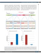
Figure 4. Sanger sequencing of the edited region of the PRDM1 locus. . (A) The diagram shows the expected homology directed repair (HDR) template inserted into the PRDM1 locus. (B) The DsRed reading frame, followed by the SV40 stop signal, was inserted in-frame into the PRDM1 gene locus. The enlarged boxes show the junctional sequence between each adjacent fragment. Blue and red arrows indicate the primer pair located outside of the HDR template area to amplify the genome DNA fragment for Sanger sequencing. (C, D) Relative PRDM1 transcript levels of exon 5 and exon 2 in PRDM1-/- NK cells versus wild-type NK cells were measured by quantitative reverse transcription polymerase chain reaction (RT-PCR) and normalized to RPL13A. The expression levels were calculated by the 2^-DDCt method, and PRDM1 expression level in wild-type NK cells was set at 1.0.
haematologica | 2021; 106(9)
2433
