
Page 144 - 2021_09-Haematologica-web
P. 144
G. Dong et al.
TLR4,19 CCR4,20 VEGF,21 TP63 and IRF4) were upregulated >20-fold in PRDM1-/- cells (Online Supplementary Figure S8D). TOX2 is a critical transcription factor for NK-cell mat- uration/development upstream of T-bet/TBX21, suggesting that PRDM1 may regulate NK-cell development and func- tion via TOX2.22 Similar to a previous study in mouse NK cells, there was a marked increase in the expression of IRF4 and its target genes upon PRDM1 KO.23 In contrast to the PRDM1 KO cells, the WT cells showed enrichment of genes associated with the FOXO3 pathway, TP53/63, and NK-cell-mediated cytotoxicity (Online Supplementary Figure S8E). Several gene signatures associated with endosomal sorting, lysosomes and secretion, and cell-to-cell communi- cation (e.g., E-cadherin stabilization) were enriched in these cells. Other than these, gene sets associated with quies- cence and IL-12, P38 MAPK and TNFR1 signaling were also enriched (Figure 7D). The genes that were downregulated included T-cell and NK-cell signaling and KIR3DL1-3 or KIR3DL members, which generally transduce inhibitory signals upon ligand binding (Online Supplementary Figure S8F). A recent report indicated that several immune check- point molecules in CD8+ T cells,24 including LAG-3, are upregulated by PRDM1 alone or in combination with MAF. We examined our data and found that the co-inhibitory receptors/molecules LAG3, LILRB1, LILRB3, and CD244 were downregulated in PRDM1-/- cells, which may thus impair immune checkpoints in NK cells similarly to cyto- toxic T-cells.
We performed qRT-PCR on selected transcripts based on their significant alteration on RNA-sequencing and their potentially important biological functions, comparing LAG3, GNLY, PRF1, TOX2 and CCR4 expression in bulk donor #2 NK PRDM1-/- cells versus donor #2 NK WT cells.
The qRT-PCR results were concordant with our RNA- sequencing results with higher levels of TOX2 and CCR4 and lower levels of LAG3, GNLY and PRF1 in PRDM1-/- cells (Online Supplementary Figure S8G). Furthermore, flow analy- sis indicated a concordant decrease in LAG3 protein expres- sion in NK PRDM1-/- cells (Online Supplementary Figure S8H).
Discussion
We and other groups have identified several frequent mutations in ENKTCL, including a number of potential tumor suppressor genes such as PRDM1, TP53, DDX3X and BCOR.5-8,25-27 We also found that ENKTCL has a marked DNA hypermethylation phenotype with inactiva- tion of multiple tumor suppressors through this mecha- nism.5 Some of the tumor suppressor genes are inactivated by a combination of genetic and epigenetic mechanisms, including PRDM1 and DDX3X. The most common activat- ing mutations involve the JAK/STAT3 pathway, affecting ~20-30% of cases.15,16 This may be related to the critical dependence of NK cells on IL-2 or IL-15, which signal through STAT3, STAT5A and STAT5B.25 Interestingly, there is frequent DNA methylation of PTPN6 (SHP1), a negative regulator of STAT3 and NK-cell receptor activation.28-32 As PRDM1 is a commonly inactivated tumor suppressor gene in ENKTCL, and as there is good evidence that PRDM1 reg- ulates normal NK-cell as well as T-cell homeostasis,6,26 we therefore focused on PRDM1 deletion to elucidate the func- tions of PRDM1 in normal NK cells and how these relate to its tumor suppressor function.
The major challenge in deciphering the role of a genetic aberration in the pathogenesis of a lymphoma is the fre-
A
B
C
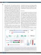
Figure 3. Schematic illustration of CRISPR/Cas9-mediated insertion of GFP or DsRed fusion tag to PRDM1 exon 5 through a homology directed repair template to induce early termination of PRDM1 expression. (A) Guide RNA sgRNA2 sequence and schematic gene structure of PRDM1. Light green boxes and numbers rep- resent the exons of the PRDM1 gene. The sequence of guide RNA sgRNA2 used for the homology directed repair (HDR) template-mediated CRISPR/Cas9 is shown above. (B) HDR templates were assembled with fluorescent tag GFP or DsRed open reading frame and a SV40 virus transcriptional stop signal (red box) in exon 5. (C) Western blot analysis of PRDM1 protein expression in PRDM1-/- NK cells and wild-type control cells (left panel). PRDM1 target gene MYC expression was meas- ured by quantitative reverse transcription polymerase chain reaction (right panel). Target gene normalization and relative expression levels were determined as described in Figure 2.
2432
haematologica | 2021; 106(9)
