
Page 143 - 2021_09-Haematologica-web
P. 143
Functional and oncogenic roles of PRDM1 in NK cells
A
C
B
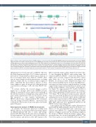
Figure 2. Sanger sequencing demonstrating the PRDM1 deletion in clones #3 and #5. (A) Guide RNA sgRNA4 sequence and schematic gene structure of PRDM1. Light green boxes and numbers represent the exons of the PRDM1 gene. (B) TOPO cloning showed that both clones #3 and #5 harbored the same bi-allelic deletion, a 79-bp deletion in one allele (comprising 35 bp from exon 4 [411-445] and 44 bp from the adjacent intron 3 and a 11-bp deletion in the other allele (413-424) (Ref: NM_001198). (C) Western blot analysis of PRDM1 protein in clones #3 and #5 (upper panel). Parental wild-type natural killer (NK) cells were used as a positive con- trol. NK lymphoma cell lines, NKYS and KAI3, were used as positive and negative controls, respectively. PRDM1 target gene MYC expression of PRDM1-/- clone #5 versus wild-type NK cells was measured by quantitative reverse transcription polymerase chain reaction (qRT-PCR) (lower panel). The target gene expression levels were normalized to RPL13A, and relative expression was calculated using the 2^-DDCt method. The expression of MYC in WT NK cells was set at 1.0.
GFP plasmid vector based approach18 on PRDM1-/- NK clone #3 (Online Supplementary Figures S5-S7). All the double mod- ified cells we obtained harbored heterozygous deletions of the second targeted gene, TP53, DDX3X or PTPN6. There were no major changes in cellular characteristics or cloning efficiency despite the loss of additional tumor suppressor genes, suggesting that heterozygous deletion of these tumor suppressor genes did not provide additional growth or proliferation advantage to NK cells with PRDM1 dele- tion.
To evaluate whether the observed changes could be largely due to off-target effects from CRISPR/Cas9 gene editing, we performed custom capture and sequencing of known driver mutations in lymphoma using a panel of 334 genes (Online Supplementary Table S5) but did not observe any mutations or copy number abnormalities in PRDM1-edited clones compared with the WT counterpart. Thus, the changes observed in PRDM1-/- cells were not due to alterations of the exomes of any of the genes tested in the panel (334 genes).
Gene expression analysis of PRDM1 knockout cells
To elucidate the functional alterations resulting from PRDM1 deletion, we performed RNA-sequencing analysis on PRDM1-deficient NK cells or clones and cell-age- matched WT NK cells from the same donors, including two biological replicates. We were able to detect the deletion of exon 4 sequences and insertion of the GFP sequence in the
RNA-sequencing analysis (Online Supplementary Figure S8A- C), thus disrupting the PRDM1 open reading frame. The truncated mRNA in PRDM1-/- clones were transcribed at higher levels (>2-fold) compared with their WT counter- parts, likely due to loss of negative autoregulation by PRDM1. As shown in Online Supplementary Figure S8A, these truncated mRNA did not result in any PRDM1 pro- tein expression. Initial analysis using hierarchical clustering suggested that clusters were partly driven by distinct donor profiles and that PRDM1-/- clones tended to form tight clus- ters (Figure 7A). Approximately 30% (3,684 of 12,633) of transcripts were differentially expressed (1,419 downregu- lated, 1,498 upregulated; P<0.05 and false discovery rate <0.3) (Figure 7B). As anticipated, numerous genes and path- ways associated with proliferation and cell cycle regulation and progression (e.g., cell cycle control, chromosomal repli- cation, centrosome maturation, RNA splicing, DNA repair, S-phase and G2/M transition targets, E2F targets, MYC, IRF4, E2F3 induction) were upregulated/enriched in the PRDM1-deleted clones (Figure 7C).
Genes regulated by the DREAM complex (Dimerization partner, RB-like, E2F4 and multi-vulval class B, master coor- dinator of cell cycle-dependent gene expression) were enriched upon PRDM1 loss, suggesting that the DREAM repressor complex converts into an activating complex in the absence of PRDM1. Metabolic changes, such as glyco- gen metabolism, also showed enrichment in PRDM1-/- NK cells. Various genes associated with NK-cell biology (TOX2,
haematologica | 2021; 106(9)
2431
