
Page 142 - 2021_09-Haematologica-web
P. 142
G. Dong et al.
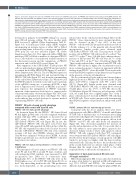
Figure 1. Schematic diagram of knock-out of PRDM1 in NK cells by the CRISPR/Cas9 system. (A) PRDM1 was knocked out by using a Cas9-sgRNA plasmid. The plasmid with both Cas9-GFP and sgRNA is shown. This approach requires cloning of GFP+ NK cells on irradiated feeder cells. Genome editing was evaluated by a high resolution melting-polymerase chain reaction assay and the PRDM1 knock-out was confirmed by western blot and Sanger sequencing. (B) PRDM1 was knocked out by electroporation of the Cas9/sgRNA RNP complex plus a homology directed repair (HDR) template with fluorescence protein gene. Double-stranded HDR DNA templates with inserted GFP or DsRed open reading frame and about 300 bp homologous sequence of exon 5 at each side of the CRISPR/Cas9 cutting site of sgRNA2 were prepared. FACS for GFP and DsRed double positive cells identified PRDM1 knock-out cells, which were confirmed by western blot and Sanger sequenc- ing. Cas9: CRISPR-associated protein 9; GFP: green fluorescent protein; NK: natural killer; PBMC: peripheral blood mononuclear cells; NHEJ: non-homologous end joining; CRISPR: clustered regularly interspaced short palindromic repeat; FACS: fluorescence activated cell sorting; PCR: polymerase chain reaction; KO: knock-out; HDRT: homology directed repair template; RNP: ribonucleoprotein.
developed for primary T-cell CRISPR editing17 for our pri- mary NK-cell genome editing. We chose another guide RNA for exon 5, sgRNA2 (Online Supplementary Table S1, Figure 3A). A homologous DNA repair (HDR) template encompassing an in-frame fusion of either GFP or DsRed open reading frame followed by a strong stop signal from SV40 poly (A) tail was included (Figure 3B, Online Supplementary Figure S1). This approach utilized HDR that introduced GFP or DsRed as markers of a disrupted PRDM1 locus. Cells with two colors indicated bi-allelic insertion of the fluorescent protein and the termination of PRDM1 expression and could be FACS-sorted (Figure 1B).
After expansion of the GFP+/DsRed+ double-positive NK cells, we performed genotyping, qRT-PCR and western blot to confirm the bi-allelic PRDM1 KO (Figures 3C and 4A-C, Online Supplementary Table S4). The low PRDM1 expression remaining in qRT-PCR (Figure 4C) and western blotting of PRDM1-/- NK cells (Figure 3C) was from the contaminating feeder cells (Online Supplementary Figure S4). When we used the PRDM1 primers located in exon 2 and exon 3, upstream of the sgRNA2 cutting site, to perform qRT-PCR, a high level of expression was observed. As PRDM1 is an autolo- gous repressor, the upregulation of PRDM1 transcripts upstream of the truncation should not be a surprise and is consistent with earlier observations (Figure 4D). MYC tran- scription was upregulated in these cells (Figure 3C). These results indicate that we had developed a separate method of generating PRDM1-/- NK cells.
PRDM1-/- NK cells showed growth advantage compared with normal wild type NK cells PRDM1-/- NK cells had a growth advantage
As shown in Figure 5A, the PRDM1-/- NK clones #3 and #5 had higher growth rates (>2 fold) compared with their normal wild-type (WT) counterparts as measured by an MTS assay. A similar effect was observed with GFP+/DsRed+ PRDM1-/- NK cells (Figure 5B). In fact, the GFP+/DsRed+ PRDM1-/- NK cells grew even better than clones #3 and #5 and expanded 4.2-fold in 6 days, while the WT NK cells expanded less than 2-fold. It must be noted that the GFP+/DsRed+ PRDM1-/- NK cells were relatively younger (~20 days after CRISPR), whereas clone #3 and clone #5 were examined ~90 days after CRISPR. A steady decline in growth rate was observed in PRDM1-/- cells after long-term in vitro culture.
PRDM1-/- NK cells had an increased fraction of cells in the S/G2M phase
We performed cell cycle analysis on PRDM1-/- NK cells (clone #3 and clone #5) at around 90 days of culture, and cells undergoing DNA synthesis were measured by EdU incorporation into DNA for 5 consecutive days. Remarkably, 40% of the PRDM1-/- NK cells were in the S/G2M phase versus ~4% in WT NK cells (Figure 5C, D) on the 3rd day after adding fresh feeder cells. This proliferation was feeder-dependent, as the proliferating rates were
reduced after feeder cells had all died (Figure 5D), but the PRDM1-/- clones demonstrated a more sustained prolifera- tion. As observed on the 7th day, >20% of the total PRDM1-/- cells showed EdU incorporation (decreased about 1.5-fold), whereas 1% of the parental cells showed EdU incorporation. Similar results were obtained with GFP+/DsRed+ PRDM1-/- NK cells. The proportion of EdU- positive cells in PRDM1-/- NK cells was 44.2% on the 3rd day and 17.8% on the 7th day (~2.5-fold drop), whereas the pro- portions of EdU-positive WT NK cells were 23.7% on the 3rd day and 4.75% on the 7th day (~5-fold drop) (Figure 5E). These results indicated that, compared with WT NK cells, PRDM1-/- NK cells had a higher rate of proliferation and a more sustained response to feeders, in agreement with the findings from PRDM1-/- clones #3 and #5. These results indi- cated that PRDM1 regulates cell proliferation and longevity in the presence of feeder cell stimulation.
We also performed cell cycle analysis and observed that a higher proportion of PRDM1-/- NK cells were in the S/G2M phase (22.9~28.5%) compared with WT control cells (18.7%) (Figure 6D). Similar results were obtained with GFP+/DsRed+ PRDM1-/- NK cells, of which 26.4% were in S/G2M phase versus the 17.0% of WT NK cells in the S/G2M phase (Figure 5F). Moreover, in both groups of NK cells, we found there was a slightly higher ratio of the S phase cell fractions (1.5~2.2-fold) in PRDM1-/- NK cells than in WT NK cells. These results unequivocally demonstrated that PRDM1 regulates cell proliferation and growth in NK cells.
PRDM1-/- primary NK cells had fewer apoptotic cells
Consistent with the above analysis, there were fewer early apoptotic cells (Q3) in PRDM1-/- clones #3 and #5 than WT NK cells at different days of observation after adding fresh feeder cells (Figure 6A). Statistical analysis showed the general trend of increase of early apoptotic cells in both PRDM1-/- and WT NK cells after feeder cell stimulation (Figure 6B). This observation paralleled the decrease in growth and proliferation of the cells. It is possible that as the cells entered a more quiescent phase, apoptosis also decreased. Similar results of fewer early apoptotic cells in PRDM1-/- NK cells compared with their WT counterpart were obtained with GFP+/DsRed+ PRDM1-/- NK cells (Figure 6C). However, the percentages of early apoptotic cells (Q3) tended to get lower, not higher, with the days in culture with feeder cells. This may be due to the fact that the cells in the second batch were at a relative earlier phase of their lifespan than the single clones #3 and #5, so the cells responded better to the feeder cell stimulation.
Generation of dual-gene knockout NK cells and off-target evaluation
Other than single-gene KO, we also introduced deletions in genes often deleted or mutated in NK-cell lymphomas (e.g. TP53, DDX3X and PTPN6). We obtained several different dual-gene KO NK cells using the pSpCas9(BB)-2A-
2430
haematologica | 2021; 106(9)
