
Page 148 - 2021_09-Haematologica-web
P. 148
G. Dong et al.
AB
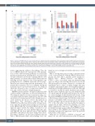
Figure 6. Apoptosis of PRDM1-/- NK cells versus wild-type NK cells. Apoptotic cells were analyzed with annexin V/propidium iodide (or DAPI) staining and a flow cytom- etry assay. Numbers in Q3 (percentage of APC or Alexa Fluor 647 annexin V-positive and propidium iodide- or DAPI-negative cells) were considered the early apoptotic cells. FITC anti-CD56 antibody staining was used to identify NK cells. Each staining was performed in duplicate. (A) Flow analysis of clones #3 and #5 from NK cells (donor #1) on day 3 and day 7 after addition of feeder cells. (B) Statistical analysis of the average percentage. (C) Experiments were repeated on NK cells (donor #2) edited by Cas9/sgRNA RNP plus homology directed repair. GFP positivity for PRDM1-/- cells and FITC CD56 positivity to identify NK cells.
C
results, supporting the validity of the findings. Thus, the loss of PRDM1 altered the transcriptome with upregula- tion of MYC, MYB and many pathways associated with growth and proliferation, including those associated with cytokine stimulation and receptor signaling. On the other hand, pathways associated with normal cellular functions including cytotoxic functions were down-regulated, sug- gesting that the loss of PRDM1 shifted the cell toward pro- liferation and survival rather than the performance of its normal effector function. The loss of immune checkpoint molecules may be more relevant in the in vivo setting, in which the cells may be able to escape from extrinsic con- trols, and could be relevant to lymphomagenesis.
CRISPR/Cas9 may generate off-target modifications that could potentially compromise our data and interpretation. We examined a number of clones for mutations using a cus- tom capture platform (Online Supplementary Table S5) with an extensive set of genes known to harbor lymphoma-asso- ciated mutations and did not observe any mutations in these genes. The observation suggested that the CRISPR/Cas9 modifications did not induce off-target muta- tions of known oncogenic drivers and that our observations in the PRDM1 KO cells were likely PRDM1-specific. Our approach cannot exclude the editing of genetic loci not examined. Therefore, we used a different approach to gen- erate KO NK cells from different donors, with different guide RNA, and without subsequent cloning. Since off-tar- get editing is unlikely to be the same in different cells and with a different guide RNA, critical findings were con-
firmed in our second approach with modification of a bulk population.
One of our long-term goals is to study cooperative effects of two or more mutations. With the PRDM1-deleted back- ground, we were able to generate double mutant (i.e. PRDM1-/-/TP53+/-, PRDM1-/-/DDX3X+/-, and PRDM1-/-/ PTPN6+/-) clones successfully using the plasmid/cloning approach. The growth of the NK cells started to slow down after prolonged in vitro culture. Therefore, to accelerate the experiments and to reduce secondary changes that may accumulate in prolonged culture, we tested the feasibility of simultaneously editing multiple genes in NK cells. Although it is feasible to modify two genes simultaneously, modify- ing three genes simultaneously was very inefficient using our current approach, probably partly because the cell can- not tolerate multiple double-stranded DNA breaks. Using the plasmid CRISPR/Cas9 KO strategy, we have only isolat- ed heterozygous deleted clones, but with the current, more efficient Cas9/sgRNA RNP electroporation procedure, besides inserting a fluorescent protein gene in one allele, 70% of the other alleles were also modified by indels as analyzed by an online software ICE (Synthego, CA, USA) (Online Supplementary Figure S10). This suggested a more promising homozygous gene editing by the RNP electropo- ration method. The CRISPR/Cas9 technology is rapidly advancing, and innovative approaches can be incorporated in the future as they appear.
While the loss of function of PRDM1 is frequent5,6 and likely to be one of the early alterations in the development
2436
haematologica | 2021; 106(9)
