
Page 106 - 2021_09-Haematologica-web
P. 106
I. Veletic et al.
confounding factors for overall survival in this cohort of patients. We therefore performed a multivariate analysis of survival after adjusting the model for these four predictors (Figure 7B). This analysis confirmed that PD1-overexpressing fractions of CD8+ cells, but not CD4+ cells, before initiation of ruxolitinib treat- ment, independently predicted overall survival (hazard ratio: 2.48; P=0.03). Taken together, our results demonstrated that increased PD1+/CD8+ T-cell subsets were significantly associated with a high risk of death in ruxolitinib-treated MF patients.
Discussion
In the current study, we found that T-cell subsets of patients with MF shifted from a quiescent to an activated state and that treatment with ruxolitinib reduced the activation of both helper CD4 and cytotoxic CD8 T cells in a time-dependent manner (Figure 8). The activation pattern of CD8+ T cells was significantly similar to that of polycythemia vera,31 including decreased TN and TCM, unaltered TEM and increased TEFF subsets. However, in MF, CD4+
cells were also considerably skewed toward an effector cell pheno- type, unlike polycythemia vera. Whereas CD8+ cells are activated by major histocompatibility complex (MHC) type I molecules, priming of CD4+ T cells is restricted to MHC class II on predomi- nantly monocyte-derived antigen-presenting cells. Because MHC expression is induced by activated JAK2,32 our data point toward a predominant role of neoplastic monocytes in aberrant T-cell responses in MF. Furthermore, circulating monocyte-derived den- dritic cells from patients with MF were extremely efficient in anti- gen uptake as compared to dendritic cells from healthy individuals, despite their reduced numbers and function.33 In comparison, T cells of acute myeloid leukemia patients at diagnosis are predomi- nantly CD8+,34,35 whereas their activation seems to differ minimally from normal cells in both CD4+ and CD8+ subsets.36,37 Overall, the activation status of T-cell subsets in patients with MF is consistent with an ongoing antineoplastic immune response, characteristic of the “T-cell inflamed” immune signature.38
Over the last decade, broad clinical experience has been acquired in treating MF patients with ruxolitinib. Overall, decreased rates of infections and spleen reduction with ruxoli-
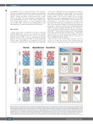
Figure 8. Schematic representation of the circulating T-cell subset repertoire in patients with myelofibrosis at baseline and after treatment with ruxolitinib. The left panel summarizes flow cytometry data from 47 patients with myelofibrosis (MF) analyzed in this study. Prior to treatment, T cells are skewed towards effector subsets (middle) and PD1-expressing T cells are increased (bottom) compared to those in 28 age-matched normal donors. In addition, disease progression shifts T-cell sub- sets towards CD8+ phenotypes (top). Ruxolitinib treatment reverts the resting:effector T-cell ratio to normal (middle), but has little effect on CD4/CD8 subsets or per- centage of PD1+ cells. The right panel summarizes correlations of differentiation subsets (top) and PD1+ fractions (bottom) with the clinical parameters at treatment baseline (dashed line). Increases in CD8/PD1-coexpressing subsets are associated with a lack of spleen response. In addition, a CD8-predominant T-cell repertoire is found in patients with monocytosis and low platelet counts, whereas abundance of PD1-overexpressing CD8+ T cells is predictive of poor overall survival.
2394
haematologica | 2021; 106(9)
