
Page 81 - 2021_06-Haematologica-web
P. 81
Hyaluronan/CD44-mediated VLA-4 activation in AML
cells confirmed the specificity of the HA-CD44 interactions in triggering adhesion on VCAM-1 expressed by stromal cells (Online Supplementary Figure S7B).
We next investigated downstream signaling of this VLA- 4/VCAM-1 interaction in AML cells by western blotting and quantified phosphorylation levels of previously shown signaling molecules that are important for AML cell sur- vival, namely Akt, ERK, IkB and mTOR.1 Primary cells from five different AML patients were treated with HA and/or with VCAM-1-coated beads, where indicated. In contrast to brief HA treatment, VCAM-1-coated beads alone were sufficient to increase phosphorylation of Akt, ERK, IkB and mTOR in primary AML cells (Figure 6A, B). This can be attributed to the experimental three-dimensional nature of this system, which allows a lot more cells to bind to the VCAM-1-coated beads than immobilized VCAM-1 used for microscopy (Online Supplementary Figure S8A). In line with this, VCAM-1 also triggered phosphorylation of ERK, IkB, FAK and paxillin (Pax) in OCI-AML3 cells (Online Supplementary Figure S8B).
tive effect of the CD44-VLA-4-dependent cell adhesion in the context of chemotherapy. We found that OCI-AML3 cells adherent to a co-immobilized substrate of HA and VCAM-1 underwent less doxorubicin-induced apoptosis than cells lacking such a substrate. CD49d expression was mandatory for the protective effect as CD49d knockdown cells were not protected by HA/VCAM-1 (Figure 6C). The importance of CD49d in leukemic progression was also confirmed by long-term in vivo engraftment experiments in NSGS mice. AML progression was decelerated upon engraftment of CD49d knockdown (shCD49d) OCI-AML3 cells as compared to engraftment of control cells (shCont) (Figure 6D). In a xenotransplant model anti-CD49d anti- body treatment altered the organ-specific localization of engrafted MOLM-13 cells, but did not significantly prolong the overall survival of mice undergoing cytarabine (AraC) treatment (Online Supplementary Figure S9). At this point we were not successful in establishing a model for testing stan- dard induction therapy (combined AraC-doxorubicin) and anti-CD49d treatment in immunodeficient mice, as doxo- rubicin requires careful further dosing studies to avoid severe toxicities.
In light of the key role of active Akt, MAPK, and NF-kB signaling in leukocyte survival, we next tested the protec-
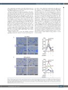
A
B
Figure 5. Hyaluronic acid treatment leads to strong interaction between acute myeloid leukemia cells and stromal cells. OCI-AML3 cells (n=4) (A) or primary cells from bone marrow (BM) aspirates from four different patients with acute myeloid leukemia (AML) (B) were pretreated or not with HA and/or αCD44 (clone 515) or αCD49d (clone HP2/1) and allowed to adhere to M2 stromal cells for 30 min. The number of AML cells that bound to the stromal cells was counted on bright field images with additional DAPI staining by fluorescence microscopy. One-way analyses of variance with multiple comparisons were used. Images were taken at 20x magnification. Bars, 20 mm. *P<0.05; **P<0.01.
haematologica | 2021; 106(8)
2109
